Hypsibius exemplaris
Hypsibius exemplaris is a tardigrade in the genus Hypsibius (Hypsibiidae, Parachela), described by Gąsiorek, Stec, Morek & Michalczyk in 2018.
Where it has been recorded
What we have read about it
Facts we extracted from the literature into our knowledge graph, each with the source and page it came from.
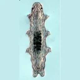
- Adults are ~500 µm long.Gabriel et al. 2007. The tardigrade Hypsibius dujardini, a new model for studying the evolution of development. Developmental biology. p547, Fig. 2 caption
- Adults have approximately 1400 ± 118 nuclei.Storage cell proliferation during somatic growth establishes that tardigrades are not eutelic organisms. Biology Open 13 (2024) bio060299 Results, nuclei counts
- The H. exemplaris genome assembly GCA_002082055.1 (2017, scaffold level) spans 104.2 Mb.NCBI Datasets genome reports for taxon Tardigrada dataset report for GCA_002082055.1
- The H. exemplaris septulum is 1.6 ± 0.2 µm long (1.3–2.0 µm; pt 6.2 ± 0.6, 4.9–7.5), n = 30.GĄsiorek et al. 2018. An integrative redescription of Hypsibius dujardini (Doyère, 1840), the nominal taxon for Hypsibioidea (Tardigrada: Eutardigrada). Zootaxa. p59, Table 5 (Hoyer's-mounted type series of H. exemplaris), row Septulum
- At 4 dph, 10 of 23 juveniles had started or completed ecdysis, leaving through the front of the old cuticle.Yamakawa & Hejnol 2024. Ecdysteroid-dependent molting in tardigrades. Current biology : CB. p. 5805, text; Figures 1E, 1F
- The predicted telomeric repeat GATGGGTTTT is strongly enriched at both ends of at least three of the five DNA Zoo Hi-C chromosomes.Coke et al. 2026. Modified meiosis in the tardigrade Hypsibius exemplaris maintains heterozygosity across the genome. Genetics. p. 6, Results; Figure 4a
- Female germ cells form branched clusters of 21–27 cells joined by cytoplasmic bridges. The corresponding figures are 8 cells in Dactylobiotus parthenogeneticus and more than 30 in Thulinius ruffoi.Jezierska et al. 2021. Oogenesis in the tardigrade Hypsibius exemplaris Gąsiorek, Stec, Morek & Michalczyk, 2018 (Eutardigrada, Hypsibiidae). Micron (Oxford, England : 1993). p. 6, §3.3; Figures 3A–3C; p. 10, §4.2 comparison
- Tardigrade-specific genes (phylostrata Tardigrada and H. exemplaris) make up 36.9% of H. exemplaris genes. Phylostrata came from reciprocal Diamond BLASTP (e < 1e-5) against 3,376 UniProt reference proteomes, one per genus.Li et al. 2024. Conserved and specific gene expression patterns in the embryonic development of tardigrades. Evol Dev 26:e12476 p. 3 of 15; Methods 5.4 (p. 12); Table S1 (every BV898 gene with phylostratum and GO terms)
- H. dujardini mixed-stage Petri stocks with glycerol added to 15% and frozen slowly to −80 °C in a closed styrofoam box could re-establish cultures after 51 and after 365 days, with roughly 50% of individuals surviving in each case.Gabriel et al. (Goldstein lab). Simple cryopreservation method for Hypsibius dujardini, from Gabriel et al. 2007 (1 p.) p1, whole text: "roughly 50% of the individuals from mixed-stage cultures surviving in each case"
- Whole-animal DAPI nucleus counts of H. exemplaris (confocal z-stacks through the entire animal, counted in Imaris) give approximately 1140 +/- 60 nuclei in hatchlings (n = 25) and approximately 1400 +/- 118 in fully-grown adults (n = 22).Quiroga-Artigas & Moriel-Carretero 2024. Storage cell proliferation during somatic growth establishes that tardigrades are not eutelic organisms. Biology open. Results, first paragraph ("approximately 1140 +/- 60 nuclei in hatchlings and approximately 1400 +/- 118 [in adults]"); Fig. 2A,B (violin plots, n = 25 and n = 22); Materials and Methods, "To count the total number of cells in hatchlings and adults, Imaris software was employed"
- Supplementary Table 1 lists the 62 H. dujardini embryos analysed, in BLIND order, with annotated times 0–2,970 min in 30-min steps (60 distinct times; 2,760 and 2,850 min occur twice). No embryo has an annotated time between 540 and 900 min.Levin et al. 2016. The mid-developmental transition and the evolution of animal body plans. Nature 531:637–641 Supplementary Information pp. 6–7, Supplementary Table 1, H. dujardini rows (HD0001–HD0126); counted by us
- After injection without electroporation, precise joining still dominated (17/22 hemerythrin, 19/22 dsup clones). The other 8/44 had 1 bp deletions, substitutions or 46–78 bp insertions at the junction, which never occurred with electroporation.Kumagai et al. 2022. Application of CRISPR/Cas9 system and the preferred no-indel end-joining repair in tardigrades. Biochemical and biophysical research communications. p. 199–200, §3.4; Figure 3; Table 1
- At the segmentation stage EIC nuclei have more diffuse DAPI staining than surrounding nuclei: the mean intensity along line scans is significantly lower (unpaired t-test, p < 0.0001; 6 embryos, 4 EIC and 4 surrounding nuclei each, 24 + 24 nuclei).Heikes et al. 2023. The embryonic origin of primordial germ cells in the tardigrade Hypsibius exemplaris. Developmental biology. p43–44 Fig. 1B–C′; Supplementary Fig. 1 (mmc3 p2); p56 §5.14
- H. exemplaris and R. varieornatus each have three extradenticle paralogues (PBC domain plus homeodomain), from duplications in a common tardigrade ancestor, and single orthologues of homothorax, Distal-less and aristaless. He-AL has a homeodomain but no OAR domain; Rv-AL has one.Game & Smith 2020. Loss of intermediate regions of perpendicular body axes contributed to miniaturization of tardigrades. Proceedings. Biological sciences. p. 2–3 (Results a); Fig. 1; Fig. S1
- The dorsal, saddle-shaped brain of the scanned H. exemplaris measures 1,392.90 µm³, 1.0% of total body volume, which the authors note is about twice the relative brain volume of honeybees and more than that of larger insects, though smaller than the 2.9–4.3% of featherwing beetles.Gross et al. 2019. X-ray imaging of a water bear offers a new look at tardigrade internal anatomy. Zoological Letters 5:14. Table 1 (brain 1392.90 µm³, 1.0% TBV); Results, 'The dorsal, saddle-shaped brain (1.0% TBV)'; Discussion, comparison with honeybees, dytiscids and ptiliids
- He-Sp6-9A and He-Sp6-9B are close neighbours on one scaffold and sister sequences in the tree, so they arose by duplication in the H. exemplaris lineage. The small predicted gene between them (OQV18300, 89 aa, no conserved domain) matches the 5′ UTR of He-Sp6-9A transcripts and is taken to be its 5′ UTR.Game & Smith 2025. An Sp6-9 ortholog is expressed in developing legs in the tardigrade Hypsibius exemplaris. Arthropod structure & development. pp. 3–5 (Results 3.2); Fig. 2; FASTA file S1
- The visual organs of H. exemplaris are paired, inverse pigment-cup ocelli of about 90 cubic micrometres each, embedded within the outer lobes of the brain. X-ray nanoCT of the whole head resolves each eye as a discrete body inside the outer brain lobe, at the level where the pharynx meets the buccal tube.Dutta et al. 2026. Extensive opsin gene expansion and non-cerebral origin of the minimalist eye in a model tardigrade. bioRxiv. bioRxiv p5, lines 151-153; Fig. 3a,b (p25 caption, lines 792-809); Supplementary Fig. 4
- RNA-seq of storage cells released by dissection (GSE212632) shows SAHS transcripts 5–20× more abundant than in the whole body, about the ratio of storage-cell number to body. In H. exemplaris without preconditioning, 6 of 10 SAHS paralogues exceed 1,000 TPM, while 11 of 12 CAHS paralogues are induced at least twofold by preconditioning.Tanaka, Aoki & Arakawa 2023. In vivo expression vector derived from anhydrobiotic tardigrade genome enables live imaging in Eutardigrada. PNAS 120:e2216739120 p. 3–4; Fig. 2C, F; Methods, RNA Sequencing
- In the three tardigrade species studied the brain is a dorsal, bilaterally symmetric ganglion bearing a central neuropil and a defined number of immunoreactive somata: about 14 serotonin-like and about 26 RFamide-like (Macrobiotus cf. harmsworthi), about 12 allatostatin-like (Paramacrobiotus richtersi) and 2 perisulfakinin-like (Hypsibius dujardini).Mayer et al. 2013. Selective neuronal staining in tardigrades and onychophorans provides insights into the evolution of segmental ganglia in panarthropods. BMC evolutionary biology. p3 right col., Results 'Organisation of the central and peripheral nervous system in the three tardigrade species studied'; Fig. 3 (p4) and its caption
- Direct MALDI-TOF tissue profiling of single dissected H. exemplaris brains (n = 15) detected He-PDF-1 and He-PDF-2 but not the predicted mass of He-PDF-3; extracts of 100 brains (n = 2) yielded all three predicted masses, with He-PDF-1 the most abundant, and Orbitrap fragmentation confirmed the amino acid sequences of all three peptides. The mature peptides are 18 amino acids long.Dutta et al. 2025. Pigment-dispersing factor neuropeptides act as multifunctional hormones and modulators in tardigrades. Open biology. Open Biology 15:240242 section 2.5, pp8-9; figure 6f,g; electronic supplementary material figure S10
- In the Heikes et al. mean lineage (nine H. exemplaris embryos), cell cycles lengthen with generation: mean 45, 44, 44, 54, 72 and 111 min for generations 1-6 (the time from a cell's birth to its division), with 3-10 min spread between cells of one generation. The daughters of AVPp and PVAa that don't go inside (AVPpa, PVAap) take ~102 min, ~30 min longer than their cousins; AVPpp and PVAaa take 181-201 min.kiraheikes/ELM example data: mean division time of every cell to the 128-cell stage in nine H. exemplaris embryos, with SD across embryos (Heikes et al. 2023 Fig. 8B data) EXAMPLE_DATA sheet 1, BranchLength and ErrorBars; cycle = cell division time minus parent's; computed 18 September 2026; Heikes et al. 2023. The embryonic origin of primordial germ cells in the tardigrade Hypsibius exemplaris. Developmental biology. Fig. 8B (the same data)
- In Yoshida 2017's H. exemplaris egg RNA-seq, the largest change between consecutive days is day 2 to day 3 (1 − Pearson r of mean log-CPM profiles 0.267), against 0.057 (day 1→2), 0.170 (day 3→4) and 0.080 (day 4→5), with 0.028-0.041 between replicates of one day. This independent dataset puts the major developmental transition between ~48 and ~72 h, where Levin 2016's mid-developmental transition falls on its corrected ~98 h axis (~47-53 h).Yoshida et al. 2017. Comparative genomics of the tardigrades Hypsibius dujardini and Ramazzottius varieornatus. PLoS biology. egg day 1-5 RNA-seq (GSM2472465-79), mean log CPM per day over all genes with variance; computed 18 September 2026
- Each of the four trunk segments has one ventral ganglion, a dense cluster of cell bodies without beta-tubulin signal, across which the paired ventral nerve cords swell to roughly three times the width of the connectives between ganglia and are joined by fibrous commissures. In the measured specimen the maximum widths of ganglia 1 to 4 were 12.4, 12.8, 13.4 and 8.0 micrometres: the first three are alike and the fourth is much smaller. Nerves run from each ganglion into the legs of its segment, and leg ganglia are visible within the legs.Smith & Jockusch 2014. The metameric pattern of Hypsibius dujardini(Eutardigrada) and its relationship to that of other panarthropods. Frontiers in Zoology. Frontiers in Zoology 11:66, pp3-4, 'Nervous system anatomy'; Figs 3A,E-G
- The adult mouth is surrounded by six peribuccal lobes.Gross et al. 2017. External morphogenesis of the tardigrade Hypsibius dujardini as revealed by scanning electron microscopy. Journal of morphology. Fig. 2g caption (p567) 'six peribuccal lobes (separated by dotted lines)'
Showing 24 of 573, taking the best-supported facts from each source in turn. Much of the rest is on the embryo, genomics and anatomy pages.
Open questions
- Are the primordial germ cells of H. exemplaris specified by induction, by inherited germ plasm, or by a mix of both, and which modes occur across Tardigrada?Posed by Heikes et al. 2023. The embryonic origin of primordial germ cells in the tardigrade Hypsibius exemplaris. Developmental biology. p. 52: "It remains an open question what modes of PGC specification exist in the tardigrade phylum." … "Future studies are needed to clarify the mode of PGC specification in this system."
- How many neural progenitor cells build the brain in tardigrade embryos (T. stephaniae; H. exemplaris), and when do they delaminate relative to the ganglion progenitors?Posed by Hejnol & Schnabel 2005. The eutardigrade Thulinia stephaniae has an indeterminate development and the potential to regulate early blastomere ablations. Development (Cambridge, England). p. 1353: "Although we could not determine the number of cells forming the brain"
- What neuron types, transmitters and synaptic connections make up the H. exemplaris nervous system?Posed by Ana & Saul 2025. The tardigrade as an emerging model organism for systems neuroscience. arXiv (q-bio.NC). p. 3: "neuronal identities and types have not been established, their connectome has yet to be reconstructed"
- What do engrailed, Pax3/7, Wnt4, Wnt16B, hedgehog and other homeobox genes do in H. exemplaris segmentation, and does Engrailed act before segments are visible?Posed by Chavarria et al. 2021. Extensive loss of Wnt genes in Tardigrada. BMC ecology and evolution. p. 15: "The precise developmental roles of Wnt4, Wnt16B, and other Wnt genes need to be clarified" … "other commonly conserved components of the segment polarity network, such as hh, need to be investigated in tardigrades"
- Were Wnt1, Wnt6, Wnt7, Wnt8 and Wnt10 lost in the ancestor of all tardigrades or only within the lineage leading to H. exemplaris and R. varieornatus?Posed by Chavarria et al. 2021. Extensive loss of Wnt genes in Tardigrada. BMC ecology and evolution. p. 9: "whether these genes were lost in an ancestor of all tardigrades, or within crown group Tardigrada is unclear"
- In H. exemplaris embryos, does FGF signal from segmental ectoderm to the endomesoderm during pouch formation, and does BMP signalling pattern the dorsal–ventral axis of the lateral ectoderm?Posed by Heikes et al. 2024. FGF and BMP pathway genes are expressed in segmentally iterated domains during segmentation of germ layers in a tardigrade. bioRxiv. bioRxiv p. 2 (abstract): "possible FGF signaling between ectoderm and endomesoderm, and possible roles of BMP signaling in dorsal-ventral patterning of lateral ectoderm"
- When do mesodermal cells become distinct from endoderm in the endomesodermal pouches of H. exemplaris?Posed by Smith et al. 2024. Developmental and genomic insight into the origin of the tardigrade body plan. Evolution & development. PDF p 3: "It is unclear precisely when mesodermal cells differentiate in H. exemplaris."
- Do transverse commissures form, even transiently, in the trunk ganglia of tardigrade embryos?Posed by Zantke et al. 2008. Three-dimensional reconstruction of the central nervous system of Macrobiotus hufelandi (Eutardigrada, Parachela): implications for the phylogenetic position of Tardigrada. Zoomorphology. p. 33: "Whether segmental commissures are transiently formed during embryonic development is unknown."
- Is the arthropod labrum a derived arthropod structure, or do onychophorans or tardigrades have a homologous structure?Posed by Martin et al. 2017. The nervous and visual systems of onychophorans and tardigrades: learning about arthropod evolution from their closest relatives. Journal of comparative physiology. A, Neuroethology, sensory, neural, and behavioral physiology. p. 579: "it is still unclear whether the labrum represents a derived feature of arthropods or whether homologous structures occur in other animal groups"
- Are tardigrade pigment-cup eyes homologous to arthropod median ocelli, to onychophoran eyes, or to other bilaterian eyes, and how diverse are photoreceptors across Tardigrada?Posed by Greven 2007. Comments on the eyes of tardigrades. Arthropod structure & development. pp. 401, 406: "the significance of these eyes for the evolution of arthropod visual systems is unclear yet" … "future studies may reveal a larger structural diversity of photoreceptors in the Tardigrada with traits usable for phylogenetic interpretations"
- Do protostome (including tardigrade) neuropsin/opsin-5 homologues work as UV-sensitive receptors or as retinal photoisomerases?Posed by Hering & Mayer 2014. Analysis of the opsin repertoire in the tardigrade Hypsibius dujardini provides insights into the evolution of opsin genes in panarthropoda. Genome biology and evolution. p. 2387: "it would be interesting to know whether the protostome neuropsin/opsin-5 homologs also have a function as UV-sensitive GPCRs or rather act as retinal photoisomerases"
- What do the opsins that are enriched in H. exemplaris eggs (rhabdomeric and related lineages) do during embryogenesis?Posed by Fleming et al. 2021. New Tardigrade Opsins and Differential Expression Analyses Show Ontogenic Variation in Light Perception. Genome biology and evolution. PDF p 8: "further work would be required to determine the exact function of these new tardigrade egg rhabdomeric opsins"
- Which opsins does each H. exemplaris eye photoreceptor express, and what are the spectral sensitivities of He-c-opsin-1 and -2?Posed by Fleming et al. 2021. New Tardigrade Opsins and Differential Expression Analyses Show Ontogenic Variation in Light Perception. Genome biology and evolution. PDF p 8: "Further analysis of the Tardigrada would be required to positive confirm the lack of dual expression within the eye."
- What are the electron-dense pigment granules of the H. exemplaris eye pigment cell made of?Posed by Dutta et al. 2026. Extensive opsin gene expansion and non-cerebral origin of the minimalist eye in a model tardigrade. bioRxiv. bioRxiv p. 10, lines 296–297: "whose biochemical" … "composition remains unknown"
- How are light signals transduced in the eyes of H. exemplaris, integrated in the brain and turned into phototaxis?Posed by Ana & Saul 2025. The tardigrade as an emerging model organism for systems neuroscience. arXiv (q-bio.NC). p. 13: "the pathways by which light signals are transduced, integrated, and transformed into behavioral outputs like phototaxis remain unknown"
52 more not shown.
Genomes and sequencing
Genome assemblies
| Accession | Name | Level | Released | Scaffold N50 (bp) |
|---|---|---|---|---|
| GCA_002082055.1 | nHd_3.1 | Scaffold | 2017-04-07 | 342,180 |
Raw sequencing data
82 runs (203.4 Gb) in 9 projects: 69 RNA-Seq, 10 AMPLICON, 1 FL-cDNA, 1 miRNA-Seq, 1 WGS. Browse them in the SRA.
The Hypsibius laboratory strain used in most studies was published as H. dujardini until 2018, when it was described as H. exemplaris (Gąsiorek et al. 2018). Genomes, runs and papers filed under H. dujardini are mostly that strain; see H. exemplaris and H. dujardini.
Papers
123 works in our corpus of 5,137 tardigrade papers name it in the title or abstract (44 in the title), from 2018. This is by exact name match, so it misses papers that use an older name or only the genus.
- Ultrastructural analysis of the dehydrated tardigrade Hypsibius exemplaris unveils an anhydrobiotic-specific architecture (2020, Scientific Reports) doi
- The tardigrade Hypsibius exemplaris dramatically upregulates DNA repair pathway genes in response to ionizing radiation (2024, Current Biology) doi
- Laboratory Culture of Hypsibius exemplaris (2018, Cold Spring Harbor Protocols) doi
- Oogenesis in the tardigrade Hypsibius exemplaris Gąsiorek, Stec, Morek & Michalczyk, 2018 (Eutardigrada, Hypsibiidae) (2021, Micron) doi
- Analyses of nervous system patterning genes in the tardigrade Hypsibius exemplaris illuminate the evolution of panarthropod brains (2018, EvoDevo) doi
- Verification of Hypsibius exemplaris Gąsiorek et al., 2018 (Eutardigrada; Hypsibiidae) application in anhydrobiosis research (2022, PLoS ONE) doi
Classification
| class | Eutardigrada |
|---|---|
| order | Parachela |
| superfamily | Hypsibioidea |
| family | Hypsibiidae |
| subfamily | Hypsibiinae |
| genus | Hypsibius |
Other species of Hypsibius: H. allisoni, H. antonovae, H. choucoutiensis, H. convergens, H. conwentzii, H. dujardini, H. fuhrmanni, H. giusepperamazzotti, H. henanensis, H. hypostomus, H. iskandarovi, H. kunmingensis, H. maculatus, H. marcelli, H. microps, H. montanus, H. morikawai, H. multituberculatus, H. murrayi, H. nivalis, H. novaezeelandiae, H. pachyunguis, H. pallidus, H. pedrottii, H. pradellii, H. repentinus, H. septulatus, H. seychellensis, H. shaanxiensis, H. valentinae, H. vaskelae.
Look it up
GBIF · NCBI Taxonomy · GenBank sequences · iNaturalist · Google Scholar
Classification, authority and notes: Degma & Guidetti 2026, Actual checklist of Tardigrada species, 45th edition (CC BY 4.0), which asks users also to cite Guidetti & Bertolani 2005 (Zootaxa 845: 1–46) and Degma & Guidetti 2007 (Zootaxa 1579: 41–53).
Records: GBIF.org, accessed 2026-09-20 through the GBIF API; each record belongs to the dataset that published it. The map shows only records released under CC0 or CC BY, with coordinates and no geospatial issue flag.
Sequence data: NCBI Assembly, Nucleotide and SRA, searched September 2026.
Papers: our corpus of tardigrade literature from OpenAlex.
How this is built
- Data
- Classification, authority and notes from Degma & Guidetti's 2026 world checklist. Records from GBIF, genomes and sequencing runs from NCBI, papers from our OpenAlex corpus of tardigrade literature, photos from Wikimedia Commons and iNaturalist (CC0 or CC BY), and facts from our knowledge graph, each with its source and page. A page shows only the parts we hold for that species.
- Built with
build_species.pywrites every species page from the checklist, joining each name by exact match to the other collections. Facts are ranked by confidence and evidence, and taken from each source in turn so no single paper fills the list. The records map is drawn as SVG from thinned GBIF coordinates.- Checked
- Only exact matches to accepted names bring in GBIF records, and fossil entries were read against their papers. The pages themselves are not tested against data.
- Limits
- Exact name matching misses papers and sequences filed under older names or only the genus. GBIF identifications are as their publishers made them. A short page means we hold little yet, not that little is known.
- More
- How the whole project is built