Dactylobiotus parthenogeneticus
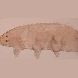
Dactylobiotus parthenogeneticus is a tardigrade in the genus Dactylobiotus (Murrayidae, Parachela), described by Bertolani in 1982.
Records
GBIF holds 59 records of it, none of them both georeferenced and openly licensed, so there is no map. See them on GBIF.
What we have read about it
Facts we extracted from the literature into our knowledge graph, each with the source and page it came from.
- The other vegetarian species have buccal tube diameters of 7–12% of tube length and stylet support insertion at 49–77% (A. granulatus 7–8%/62–65%; Minibiotus gr. intermedius 7–8%/49–54%; M. furcatus 10–12%/68–73%; Murrayon pullari and Dactylobiotus parthenogeneticus 10–12%/72–77%); Macrobiotus spp. (diet unknown) have 15–19% and 78–83%.Guidetti et al. 2012. Form and function of the feeding apparatus in Eutardigrada (Tardigrada). Zoomorphology. p129, Table 2
- Total fibres in legs I–IV (ventral + lateral + dorsal groups): Milnesium cf. tardigradum 12, 13, 13, 4; Acutuncus antarcticus 14, 13, 13, 5; Bertolanius volubilis 11, 11, 12, 6; Paramacrobiotus richtersi and Dactylobiotus parthenogeneticus 10, 11, 12, 5; Echiniscus testudo 9, 10, 11, 4. Only the ventral group was resolved in Batillipes bullacaudatus (5, 6, 6, 3).Marchioro et al. 2013. Somatic musculature of Tardigrada: phylogenetic signal and metameric patterns. Zoological Journal of the Linnean Society. Table 3 pp. 595–596, rows 'Total fibres within each leg'
- The pharyngeal cuticular lining continues into the narrow cylindrical oesophagus, and folds at the pharynx–oesophagus transition work as a valve.Guidetti et al. 2012. Form and function of the feeding apparatus in Eutardigrada (Tardigrada). Zoomorphology. p136, first paragraph; Fig. 16g
- The dorsal region of each leg receives fibres only from the dorsal group, the ventral region from all three groups. Legs I–III have similar fibre numbers (e.g. Bertolanius volubilis: three ventral, two lateral, six or seven dorsal per leg). Leg IV has only two attachment points, P1 and P2, reached by two or three fibres: one or two dorsal (to P2) and one lateral (to P1).Marchioro et al. 2013. Somatic musculature of Tardigrada: phylogenetic signal and metameric patterns. Zoological Journal of the Linnean Society. pp. 592–596, 'General remarks on leg musculature'; Table 3
- The buccal tube is a rigid cylinder whose diameter differs between species; it is straight in Milnesium cf. tardigradum and generally has an anterior-ventral bend in the parachelan species.Guidetti et al. 2012. Form and function of the feeding apparatus in Eutardigrada (Tardigrada). Zoomorphology. p130, 'Buccal tube'
- Eutardigrade muscle architecture is more complex than heterotardigrade: eutardigrade attachment points mostly serve four to nine fibres (generally more than three), whereas those of Echiniscus testudo serve one or two; E. testudo has fewer but the thickest fibres seen in tardigrades, its two dorsal strands merged for most of their length and no interlongitudinal muscles.Marchioro et al. 2013. Somatic musculature of Tardigrada: phylogenetic signal and metameric patterns. Zoological Journal of the Linnean Society. p. 584 'Tardigrade musculature'; p. 593 'Dorsal muscle group'; p. 600 Discussion
- The cuticular eutardigrade buccal-pharyngeal apparatus can be divided into four parts, buccal ring, buccal tube, stylet system and pharynx, followed by the cuticle-lined oesophagus, all part of the foregut.Guidetti et al. 2012. Form and function of the feeding apparatus in Eutardigrada (Tardigrada). Zoomorphology. p130, Results 'Anatomy of the buccal–pharyngeal apparatus'; Fig. 1a (p131)
- In eutardigrades the lateral group has two crossing longitudinal strands, one of six fibres (J-m+d, m+d–l, l-k+e, k+e-h+f, h+f-g2, g2-χ) running the whole body and one of four (H-n+c, n+c-m+d, m+d-k+e, k+e-h+f), with seven lateral nodes alternating uneven (n+c, l, i, g1+g2) and even (m+d, k+e, h+f). Echiniscus testudo has a single meandering strand of three thicker fibres.Marchioro et al. 2013. Somatic musculature of Tardigrada: phylogenetic signal and metameric patterns. Zoological Journal of the Linnean Society. pp. 589, 591, 'Lateral muscle group'; Figs 3–7; Table 2 p. 594
- In parachelans the anterior buccal tube is ventro-laterally surrounded by cuticular crests and laminae, named the buccal crown, which anchor the anterior ends of the stylet protractor muscles (M1 and M6); Milnesium lacks it.Guidetti et al. 2012. Form and function of the feeding apparatus in Eutardigrada (Tardigrada). Zoomorphology. p131 ('we propose the name “buccal crown”'); p141 Discussion 'Buccal tube'
- Seven large nodes lie on the ventral midline, uneven A, C, E, G and even B, D, F. Node A always sends two fibres to the ventral region of leg I (A-LIc, A-LId); each even node sends one fibre to the previous leg and one to the following leg, except F which lacks the forward one. A-LIc is the only fibre that crosses the midline, crossing its partner from the other side in all species.Marchioro et al. 2013. Somatic musculature of Tardigrada: phylogenetic signal and metameric patterns. Zoological Journal of the Linnean Society. pp. 583, 586, 'Ventral muscle group'; Figs 8–9; Table 3
- The buccal tube has four oval perforated areas with many ~0.1 µm holes, symmetrically on each side of the stylet sheaths (one dorsal and one ventral per side), through which sensory processes of the buccal sensory organs pass.Guidetti et al. 2012. Form and function of the feeding apparatus in Eutardigrada (Tardigrada). Zoomorphology. p131–132, 'oval perforated areas'; Figs 5d, e, 8e, 9e; p141 function
- In every species studied two ventral longitudinal strands per side (inner and outer) run from the head to leg IV. In eutardigrades each strand has six fibres (outer a-J, J-K, K-L, L-M, M-N, N-O; inner H*-H, H-K, K-L, L-M, M-N, N-O); in parachelans the strands are fused between O and K, in Milnesium cf. tardigradum only between K–L and M–N. Echiniscus testudo has four fibres per strand, fused between K and L.Marchioro et al. 2013. Somatic musculature of Tardigrada: phylogenetic signal and metameric patterns. Zoological Journal of the Linnean Society. pp. 583, 586–587, 'Ventral muscle group'; Table 2 p. 594; Fig. 8
- In the parachelan species the stylet sheaths lie ventral to the buccal tube and the stylet furcae lateral to it, and the long piercing stylet (sword-shaped anterior tip, enlarged posterior end jammed in the furca) runs in a furrow between the ventral lamina and the ventral wall of the buccal tube.Guidetti et al. 2012. Form and function of the feeding apparatus in Eutardigrada (Tardigrada). Zoomorphology. p132–133, 'Stylet system'; Figs 7b, d, 9a, 10b, c, 12b, 14a
- Serial homologies give a clear metameric pattern in all three muscle groups. Ventral group: four sets of serially homologous points (A, C, E, G; B, D, F; L, M, O; H, LIa, LIIa, LIIIa). Lateral group: two (even h+f, k+e, m+d; uneven n+c, l, i, g1+g2). Eutardigrade dorsal group: three (7, 11, ηa; 6, 10, λ; 5+4, 8+9, 12+13). Body segments 3 and 4 (legs II and III, counting a one-segment head) show the strongest pattern; the head and segment 5 (leg IV) the weakest.Marchioro et al. 2013. Somatic musculature of Tardigrada: phylogenetic signal and metameric patterns. Zoological Journal of the Linnean Society. pp. 596–597, 'Metameric patterns'; p. 600 'Musculature metamerism in tardigrades'; Figs 6–8, 12
- The buccal tube ends inside a myoepithelial pharynx whose tripartite lumen has a thin cuticular lining; except in Milnesium, the lining bears three pharyngeal apophyses alternating with three rows of symmetrical placoids (2–3 macroplacoids and at most one, smallest and most caudal, microplacoid per row).Guidetti et al. 2012. Form and function of the feeding apparatus in Eutardigrada (Tardigrada). Zoomorphology. p135, 'Pharyngeal structures and esophagus'; Fig. 1a (p131)
- Tardigrade somatic muscles form three almost independent groups, ventral, lateral and dorsal, each of longitudinal strands plus transverse muscles; circular muscles are absent. Dorsal and lateral groups lie right under the epidermis, the ventral group over the nerve cords and ganglia. The groups meet only anteriorly at ventral node J, posteriorly at dorsal point χ, and inside the legs; transverse fibres β–J and 1–J link dorsal and ventral groups in all species studied.Marchioro et al. 2013. Somatic musculature of Tardigrada: phylogenetic signal and metameric patterns. Zoological Journal of the Linnean Society. pp. 583–584, Results 'Tardigrade musculature'; p. 592 'Dorsal muscle group'; Figs 3–7
Showing 16 of 24, taking the best-supported facts from each source in turn.
Genomes and sequencing
Raw sequencing data
1 runs (20.9 Gb) in 1 projects: 1 Hi-C. Browse them in the SRA.
Papers
6 works in our corpus of 5,137 tardigrade papers name it in the title or abstract (4 in the title), from 2002. This is by exact name match, so it misses papers that use an older name or only the genus.
- Germ cell cluster organization and oogenesis in the tardigrade Dactylobiotus parthenogeneticus Bertolani, 1982 (Eutardigrada, Murrayidae) (2014, PROTOPLASMA) doi
- First record of Dactylobiotus parthenogeneticus Bertolani, 1982 (Eutardigrada: Murrayidae) in Mexico (2015, Check List) doi
- Oogenesis in Dactylobiotus parthenogeneticus Bertolani, 1982 (Tardigrada, Eutardigrada, Murrayidae) (2014, Acta Biologica Cracoviensia. Series Zoologia)
- Morphological and molecular identification of freshwater eutardigrade Dactylobiotus parthenogeneticus (Bertolani, 1982) in the Greater Zab River of Kurdistan Region-Iraq (2024, Cellular and Molecular Biology) doi
- Diversidad y distribución de las especies del phylum Tardigrada en la Península Ibérica, Islas Baleares e Islas Chafarinas (2002, Graellsia) doi
- Identification of the trehalose-6-phosphate synthase (tps) gene in desiccation tolerant and intolerant tardigrades (2012, The European Zoological Journal) doi
Classification
| class | Eutardigrada |
|---|---|
| order | Parachela |
| superfamily | Macrobiotoidea |
| family | Murrayidae |
| genus | Dactylobiotus |
Other species of Dactylobiotus: D. ambiguus, D. ampullaceus, D. aquatilis, D. caldarellai, D. dervizi, D. dispar, D. grandipes, D. haplonyx, D. henanensis, D. kansae, D. lombardoi, D. luci, D. macronyx, D. octavi, D. ovimutans, D. selenicus, D. taiwanensis, D. vulcanus.
Look it up
GBIF · NCBI Taxonomy · GenBank sequences · iNaturalist · Google Scholar
Classification, authority and notes: Degma & Guidetti 2026, Actual checklist of Tardigrada species, 45th edition (CC BY 4.0), which asks users also to cite Guidetti & Bertolani 2005 (Zootaxa 845: 1–46) and Degma & Guidetti 2007 (Zootaxa 1579: 41–53).
Records: GBIF.org, accessed 2026-09-20 through the GBIF API; each record belongs to the dataset that published it. The map shows only records released under CC0 or CC BY, with coordinates and no geospatial issue flag.
Sequence data: NCBI Assembly, Nucleotide and SRA, searched September 2026.
Papers: our corpus of tardigrade literature from OpenAlex.
How this is built
- Data
- Classification, authority and notes from Degma & Guidetti's 2026 world checklist. Records from GBIF, genomes and sequencing runs from NCBI, papers from our OpenAlex corpus of tardigrade literature, photos from Wikimedia Commons and iNaturalist (CC0 or CC BY), and facts from our knowledge graph, each with its source and page. A page shows only the parts we hold for that species.
- Built with
build_species.pywrites every species page from the checklist, joining each name by exact match to the other collections. Facts are ranked by confidence and evidence, and taken from each source in turn so no single paper fills the list. The records map is drawn as SVG from thinned GBIF coordinates.- Checked
- Only exact matches to accepted names bring in GBIF records, and fossil entries were read against their papers. The pages themselves are not tested against data.
- Limits
- Exact name matching misses papers and sequences filed under older names or only the genus. GBIF identifications are as their publishers made them. A short page means we hold little yet, not that little is known.
- More
- How the whole project is built