Actinarctus doryphorus
Actinarctus doryphorus is a tardigrade in the genus Actinarctus (Tanarctidae, Arthrotardigrada), described by Schulz in 1935.
Where it has been recorded
What we have read about it
Facts we extracted from the literature into our knowledge graph, each with the source and page it came from.
- Several conspicuous cells ('amoebocytes') lie ventral to the brain lobes in the A. doryphorus head.Persson et al. 2014. Brain anatomy of the marine tardigrade Actinarctus doryphorus (Arthrotardigrada). J Morphol 275:173-190. p176, p180; Figs 1B, 6-9
- Gross et al. find no support for a circumpharyngeal, cycloneuralian-like brain in tardigrades, for two reasons: there is no anterior-to-posterior perikarya-neuropil-perikarya organisation, and the neuropil region in the head does not form a ring but is restricted to the dorsal and dorsolateral space. The same restriction of the synapsin signal to a dorsal central neuropil was found in the arthrotardigrade Actinarctus doryphorus ocellatus. They note that no synapsin data exist from cycloneuralians for comparison, and that if synapsin turned out to be restricted to the dorsal region of cycloneuralian brains too, the interpretation of the whole cycloneuralian brain would need revisiting.Gross et al. 2021. Organization of the central nervous system and innervation of cephalic sensory structures in the water bear Echiniscus testudo (Tardigrada: Heterotardigrada) revisited. Journal of morphology. J Morphol 282, section 4.2, pp1307-1309
- The tardigrade median brain lobe is paired (Persson et al. 2012, 2014).Martin et al. 2017. The nervous and visual systems of onychophorans and tardigrades: learning about arthropod evolution from their closest relatives. Journal of comparative physiology. A, Neuroethology, sensory, neural, and behavioral physiology. p574 left col.
- Parasitism and commensalism were known only in marine species: Tetrakentron synaptae (100–200 µm, flat) lives only on the peribuccal tentacles of the holothurian Leptosynapta galliennei, two or three per host; Pleocola limnoriae on the isopod Limnoria lignorum (also found free); Echiniscoides sigismundi once on Mytilus mantle; Actinarctus doryphorus perhaps a facultative parasite of Echinocyamus pusillus (disputed).Ramazzotti & Maucci 1983. Il Phylum Tardigrada, 3rd edition (English translation by C. W. Beasley, 1995: The Phylum Tardigrada). Memorie dell'Istituto Italiano di Idrobiologia 41; translation published by the translator, McMurry University, Abilene, Texas. pp. 47–48 (PDF pp. 47–48): "In general there are two or three Tetrakentron on each holothurian, moving about slowly, keeping a tight grasp on their host with their claws."
- Smith & Jockusch propose that the horseshoe-shaped ventral brain lobe of H. dujardini is directly homologous both to the subesophageal ganglion and to the paired ventral lobes described for Halobiotus crispae by Persson et al. 2012: the ventral body of the horseshoe lies where the subesophageal ganglion is reported, and its branches lie where the paired ventral lobes are. They extend the same reading to the subesophageal ganglion and third brain lobe of the heterotardigrade Actinarctus doryphorus, and conclude that the ancestor of Eutardigrada, and probably of Tardigrada, had outer, inner and ventral brain lobes. They did not detect a discrete subesophageal ganglion.Smith & Jockusch 2014. The metameric pattern of Hypsibius dujardini(Eutardigrada) and its relationship to that of other panarthropods. Frontiers in Zoology. Frontiers in Zoology 11:66, pp11-12, Discussion 'Nervous system anatomy of H. dujardini and its relationship to that of other tardigrade species'
- Trunk ganglion 1 of A. doryphorus has one commissure and hemiganglia further apart than in H. crispae, where ganglia 1-3 each have two commissures; each hemiganglion has a neuropil and cell cortex.Persson et al. 2014. Brain anatomy of the marine tardigrade Actinarctus doryphorus (Arthrotardigrada). J Morphol 275:173-190. p176; p188; Figs 3, 5D, 12B
- A subpharyngeal ganglion lies ventrally in the tardigrade head (Marcus 1929; Persson et al. 2012, 2014).Martin et al. 2017. The nervous and visual systems of onychophorans and tardigrades: learning about arthropod evolution from their closest relatives. Journal of comparative physiology. A, Neuroethology, sensory, neural, and behavioral physiology. p574 right col.
- Each of the three brain lobes of A. doryphorus contains a neuropil and one commissure, with synapses seen by TEM; the authors stress neuropils because they occur in arthropod brains and had been deemed missing in tardigrade brains (Zantke et al. 2008).Persson et al. 2014. Brain anatomy of the marine tardigrade Actinarctus doryphorus (Arthrotardigrada). J Morphol 275:173-190. p180 Discussion
- The tardigrade inner, outer and median brain lobes are homologous to the arthropod proto-, deuto- and tritocerebrum respectively (Kristensen 1983), each lobe being a modified ganglion as shown by commissures within each lobe (Persson et al. 2012, 2014).Martin et al. 2017. The nervous and visual systems of onychophorans and tardigrades: learning about arthropod evolution from their closest relatives. Journal of comparative physiology. A, Neuroethology, sensory, neural, and behavioral physiology. p574 left col.
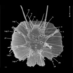
- A. doryphorus has 11 cephalic sense organs (primary and secondary clavae, internal, external and lateral cirri A, and an unpaired median cirrus; tertiary clavae absent); arthrotardigrades have up to 13, and each trunk segment up to five sensory appendages.Persson et al. 2014. Brain anatomy of the marine tardigrade Actinarctus doryphorus (Arthrotardigrada). J Morphol 275:173-190. p174 left col.; p188 left col.; Fig. 1D,E
- Serial TEM shows the A. doryphorus brain as three paired lobes in anteroposterior series: lobe 1 anterodorsal, lobe 2 anteroventral just below it (both anterior to the buccal tube), lobe 3 posteroventral, dorsal and lateral to the buccal tube; separation is more pronounced than in eutardigrades.Persson et al. 2014. Brain anatomy of the marine tardigrade Actinarctus doryphorus (Arthrotardigrada). J Morphol 275:173-190. p173 Abstract; p176, p180; Figs 6-10, 13
- Brain lobe 2 lies ventral to lobe 1, fused to its ventral part; its small anterior cluster innervates the secondary clavae and external cirri, and its larger posterior cluster contains commissure 2 and, posteriorly, a large neuropil; the outer connectives pass laterally without connecting to lobe 2.Persson et al. 2014. Brain anatomy of the marine tardigrade Actinarctus doryphorus (Arthrotardigrada). J Morphol 275:173-190. p180 TEM; Figs 7, 8, 9
- Brain lobe 3 lies dorsal and lateral to the buccal tube with dorsal and lateral clusters; the dorsal cluster holds a neuropil with at least four conspicuous elongated cells, interpreted as sensory cells for the buccal lamella (mouth cone); commissure 3 joins the lateral clusters and passes ventral to the buccal tube and stylets.Persson et al. 2014. Brain anatomy of the marine tardigrade Actinarctus doryphorus (Arthrotardigrada). J Morphol 275:173-190. p180 TEM; Fig. 10
- Brain lobe 1 has a lateral and a central cluster; the lateral cluster innervates the primary clavae and gives commissure 1, which runs through a well-defined central neuropil in the central clusters; the central cluster innervates the internal cirri (and cirrus A, CLSM); the outer connective originates from lobe 1 and runs to trunk ganglion 1.Persson et al. 2014. Brain anatomy of the marine tardigrade Actinarctus doryphorus (Arthrotardigrada). J Morphol 275:173-190. p180 TEM; Figs 4A-D, 6, 7
- A. doryphorus ocellatus were dredged at Trezen ar Skoden off Roscoff, France (45-51 m, May and July 2011) for LM (8), SEM (6) and CLSM (about 40 acetylated-tubulin + 20 tyrosinated-tubulin); eight A. doryphorus doryphorus for serial TEM came from 25 m NE of Hirsholmene, Denmark, July 1980. Animals are up to 200 um long, dorsoventrally flattened, with four toes per leg.Persson et al. 2014. Brain anatomy of the marine tardigrade Actinarctus doryphorus (Arthrotardigrada). J Morphol 275:173-190. p174 Materials and methods; p176
- The innervation of the median cirrus in A. doryphorus closely resembles nerve n13 of H. crispae, which the authors take as confirming that n13 is a rudiment of the median cirrus.Persson et al. 2014. Brain anatomy of the marine tardigrade Actinarctus doryphorus (Arthrotardigrada). J Morphol 275:173-190. p188-189
Showing 16 of 21, taking the best-supported facts from each source in turn.
Open questions
- Are the large 'one-cell' glands of arthrotardigrades homologous to insect oenocytes, and are they involved in moulting?Posed by Persson et al. 2014. Brain anatomy of the marine tardigrade Actinarctus doryphorus (Arthrotardigrada). J Morphol 275:173-190. p189 left col.: "the conspicuous 'one-cell' glands... could be an important morphological character if they were proven to be homologues to oenocytes of insects... hence it could have merit to conduct research on these cells in the future."
Genomes and sequencing
Raw sequencing data
5 runs (136.0 Gb) in 2 projects: 3 Hi-C, 2 WGS. Browse them in the SRA.
Papers
10 works in our corpus of 5,137 tardigrade papers name it in the title or abstract (4 in the title), from 1996. This is by exact name match, so it misses papers that use an older name or only the genus.
- Actinarctus doryphorus (Tanarctidae) DNA barcodes and phylogenetic reinvestigation of Arthrotardigrada with new A. doryphorus and Echiniscoididae sequences (2023, Zootaxa) doi
- Comparative myoanatomy of Tardigrada: new insights from the heterotardigrades Actinarctus doryphorus (Tanarctidae) and Echiniscoides sigismundi (Echiniscoididae) (2019, BMC Evolutionary Biology) doi
- Ultrastructure of Oogenesis in Actinarctus doryphorus (Heterotardigrada, Arthrotardigrada, Tanarctidae) (2026, ZOOLOGICAL SCIENCE) doi
- Ultrastructural studies on spermiogenesis and postcopulatory modifications of spermatozoa of Actinarctus doryphorus Schulz, 1935 (Arthrotardigrada: Halechiniscidae):Conference Information: 7th International Symposium on Tardigrada, SEP 04-07, 1997 DUSSELDORF, GERMANY (1999, Research at the University of Copenhagen (University of Copenhagen))
- Brain anatomy of the marine tardigrade actinarctus doryphorus (arthrotardigrada) (2013, Journal of Morphology) doi
- Diversity and dynamics of an interstitial Tardigrada population in the Meloria Shoals, Ligurian Sea, with a redescription ofBatillipes similis(Heterotardigrada, Batillipedidae) (1999, The European Zoological Journal) doi
Classification
| class | Heterotardigrada |
|---|---|
| order | Arthrotardigrada |
| family | Tanarctidae |
| genus | Actinarctus |
Listed as the nominotypical subspecies Actinarctus doryphorus doryphorus.
Subspecies: Actinarctus doryphorus ocellatus Renaud-Mornant, 1971.
Other species of Actinarctus: A. lyrophorus, A. neretinus, A. odissi, A. physophorus.
Look it up
GBIF · NCBI Taxonomy · GenBank sequences · iNaturalist · Google Scholar
Classification, authority and notes: Degma & Guidetti 2026, Actual checklist of Tardigrada species, 45th edition (CC BY 4.0), which asks users also to cite Guidetti & Bertolani 2005 (Zootaxa 845: 1–46) and Degma & Guidetti 2007 (Zootaxa 1579: 41–53).
Records: GBIF.org, accessed 2026-09-20 through the GBIF API; each record belongs to the dataset that published it. The map shows only records released under CC0 or CC BY, with coordinates and no geospatial issue flag.
Sequence data: NCBI Assembly, Nucleotide and SRA, searched September 2026.
Papers: our corpus of tardigrade literature from OpenAlex.
How this is built
- Data
- Classification, authority and notes from Degma & Guidetti's 2026 world checklist. Records from GBIF, genomes and sequencing runs from NCBI, papers from our OpenAlex corpus of tardigrade literature, photos from Wikimedia Commons and iNaturalist (CC0 or CC BY), and facts from our knowledge graph, each with its source and page. A page shows only the parts we hold for that species.
- Built with
build_species.pywrites every species page from the checklist, joining each name by exact match to the other collections. Facts are ranked by confidence and evidence, and taken from each source in turn so no single paper fills the list. The records map is drawn as SVG from thinned GBIF coordinates.- Checked
- Only exact matches to accepted names bring in GBIF records, and fossil entries were read against their papers. The pages themselves are not tested against data.
- Limits
- Exact name matching misses papers and sequences filed under older names or only the genus. GBIF identifications are as their publishers made them. A short page means we hold little yet, not that little is known.
- More
- How the whole project is built