A nerve cord measured, a claim killed, and an algorithm borrowed from contract theory
A long day, and an unusual number of corrections in it. We measured the tardigrade's nerve cord, which nobody had done. We published a claim about its neurons and knocked it down by lunchtime. We pulled every published statement about the tardigrade nervous system into one table and found that nearly three quarters of it was measured in a different animal from the one every laboratory uses. And we tested whether a real embryo is built the way our model builds one, with an algorithm written for designing contracts.
Four ganglia, measured
The X-ray scan published by Gross and colleagues in 2019 contains the whole ventral nerve cord, and their paper says plainly that they did not measure it. So we did. Their released label field holds the cord as one connected strand of 1,607 µm³ whose cross-section swells four times along the body. Cutting it wherever the cross-section passes 10 µm² — the connectives run at 2.5–6 µm², the swellings peak at 47–55 — gives four ganglia of 385, 430, 379 and 194 µm³ front to back, with 214 µm³ of paired connectives between them.
The fourth ganglion is half the size of the others. That matches one published count of nuclei and contradicts another, and both sit in the graph on either side of the disagreement. The cut is not delicate: any threshold from 8 to 25 µm² moves the volumes by under 10% and does not change the pattern at all.
Two more numbers fell out of the same file. Each ganglion's centre sits 4.2, 10.3 and 12.7 µm in front of its own legs' claw glands, which puts a number on the paper's own statement that each segment's ganglion lies ahead of its legs. And nervous tissue lies 2.4–3.8 µm from the traced body surface, against 5.2 µm for storage cells, 9.1 for the ovary and 11.6 for the midgut — quantifying another sentence everyone repeats, that the nervous system runs just under the skin.
The brain stays one piece, and that is a result too. We went looking for the lobes the literature describes and could not separate them: the brightness inside the brain is flat to within 15%, with no surface-to-core trend, and erosion never splits it. The largest sphere that fits inside is 2.6 µm across. So at 106 nm resolution this scan cannot tell neuropil from cell bodies — a measured limit rather than an assumption. The brain is bilaterally even to 1.7%: 688.5 µm³ left, 700.2 right.
Three labels the 2019 paper left unnamed turn out to be identifiable by registering their higher-resolution head scan onto the whole body. Label 3 is the first trunk ganglion — 86% of it falls inside the whole-body cord's first swelling — and not the separate subpharyngeal ganglion some authors report. Label 4 is the connectives running back to ganglion 2. Label 9 is a pair of outer connectives, 13 µm long, absent from the whole-body file altogether. And label 5 lies exactly where the inner connectives should run, which the paper states "were not identified". The brain page has the numbers; the glass animal now shows eleven nervous structures where it showed three.
02 · counting neuronsWe published that half the animal was nerve. It isn't
Nobody had added up the neuron counts scattered through a century of anatomy, so we did: brain, trunk ganglia, leg ganglia, the lateral and dorsal neurons, the stomatogastric cells, the extra-cerebral somata. The sum is 397–647 neurons. That is the first written-down bracket on the number, and it contains the "under 600" that the tardigrade connectome project estimates from electron microscopy.
Against the ~1,000 cells everyone quotes for this animal, 397–647 is 40–65% of the whole body — which would be the highest proportion of any animal with counted cells. We wrote that on this page in the morning.
Then we built the test instead of the press release: every animal with both a whole-body cell count and a neuron count, thirteen of them, each number with its page.
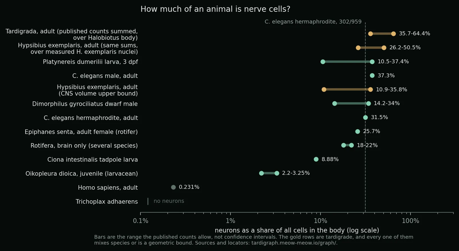
The claim fails. The highest measured ratio belongs to the Caenorhabditis elegans male — 385 neurons in 1,031 cells, 37% — and the rotifer anyone would reach for comes in at 26%. The tardigrade clears that bar only by dividing four large species' neuron counts by a small species' body. Our own scan agrees: the labelled brain and ganglia come to 2,728 µm³, which at realistic packing holds at most 123 cell bodies of 3 µm, or 417 of 2 µm — and those are upper bounds, because most of that volume is neuropil, not cells. That puts the water bear at 11–36%, straddling the worm rather than beating it.
Three things survive, and one is better than the claim we lost. The famous "about a thousand cells" for this species is an uncited aside in a 2016 genome paper, made in passing while arguing about how much bacterial DNA would show up in a sequencing run, and repeated ever since as though it were a measurement. The only real count is 1,140 nuclei in hatchlings and 1,400 in adults, and it rises as the animal grows. No tardigrade species has both counts, and no study has counted both in the same animal. And the 397–647 bracket stands on its own.
03 · the nervous system, all of itEvery published statement, in one table
This literature is spread across a century, a dozen species and several incompatible naming schemes: what one paper calls the subpharyngeal ganglion another calls trunk ganglion 1, and the abbreviations differ between figures in the same journal. Anyone building a connectome has to reconcile all of it by hand before they can start.
So we pivoted our graph by body part. The result is every published claim about each piece of the nervous system, side by side, each tied to its page or figure and to the species it was actually seen in: 104 parts, 776 statements, 42 sources, searchable.
The headline is a warning label nobody had put a number on. Almost everything done in a tardigrade laboratory uses one species — the genome, the transcriptomes, the cultures, the embryos, and the electron-microscopy connectome now being built. Only 28% of the published anatomy was seen in that animal. The rest is Halobiotus crispae (13%), Macrobiotus hufelandi (11%), Mesobiotus cf. harmsworthi (11%), Echiniscus testudo (9%) and others, spanning both tardigrade classes — in places a wider gap than mouse to human. 38 of the 104 parts have never been described in the laboratory species at all, among them the sensory cirri and clavae, the median brain ganglion, and two of the segmental nerves.
The same pivot lists 79 places where published statements directly contradict each other, both sides kept, with a note on each. Some are probably two people looking at two different animals and both being right — exactly the distinction the species tagging now makes checkable instead of guessable.
None of this is a complaint about the literature, which described whichever animal could be collected and sectioned, often decades before anyone chose a model organism. It is a label worth attaching to the anatomy anyway.
04 · six papers readHow the tardigrade eye is wired, answered
Six nervous-system papers went from sitting on the shelf to fully read and merged: 84 claims, 60 new anatomical parts, 713 links, and 12 new open questions that the papers themselves pose.
One settles a question that had been open for years. The tardigrade eye is a four-cell inverse pigment cup of about 90 µm³, sitting inside the outer lobe of the brain — and none of those four cells has an axon. Five axon-bearing neurons lie immediately beside them in the same lobe, and they are the eye's route out. Meanwhile the four cells are ringed by an epithelial protein, which reads the eye as an internalised patch of skin. So the old either/or — is the eye cerebral or epidermal? — dissolves: its position is cerebral and its derivation epidermal. Both older claims stay in the graph with the contradiction recorded.
A second paper gives the first identified-neuron map of any tardigrade: ten cells carrying a signalling peptide, and the only long-range projection ever traced in the phylum, running from the brain down the nerve cord to end as two pairs of buttons on the fourth ganglion. One of those neurites ends on the surface of the eye's photoreceptor — the first identified neuron shown to touch a tardigrade photoreceptor.
And a question posed about the armoured tardigrade Echiniscus testudo in 2013 — do the cells of the posterior head cluster serve cirrus A, the clava, or both? — was answered by the same group in 2021: two bundles to the clava, one to the cirrus. Eight years, the same animal, a better marker, and as far as we can tell nobody noticed the loop had closed.
05 · a blind testNine genes, guessed, right
Our method for putting genes on the tardigrade chromosomes is a reciprocal best-hit search: crude, fast, used across the project for markers nobody has mapped. We had no independent check on it.
At 12:41 we committed a table of nine opsin genes — the light-sensing proteins — each with the chromosome locus we had assigned it. At 12:51 a supplementary file from the eye paper arrived, naming the locus for all nine. They are the same nine. We had not seen the table; it was fetched ten minutes after our numbers went into git, and both timestamps are in the repository.
Nine out of nine is a small sample, and the honest caveat is that our method got the set right while the paper also fixes which named paralogue each one is, which the method cannot do. But it is the only blind test this route has had, and it passed.
06 · one program, two animalsAn embryo that runs a worm as well as a water bear
The embryo has a second engine: every cell owns a share of the egg, the shares fill it exactly, and no cell can overlap another. It reaches about a thousand cells in a quarter of an hour of computing, lengthens into the comma shape after 17 hours, opens a gut lumen as an empty cell, and carries signals across the faces that cells share.
Nothing in it is about water bears. A species is now either a table of divisions — each with its time, its direction and the daughters' volumes — or a set of rules in code, behind one interface. So we pointed it at Caenorhabditis elegans, whose embryos have been measured cell by cell, and asked it to predict something it had not been told: which cells touch, and how much.
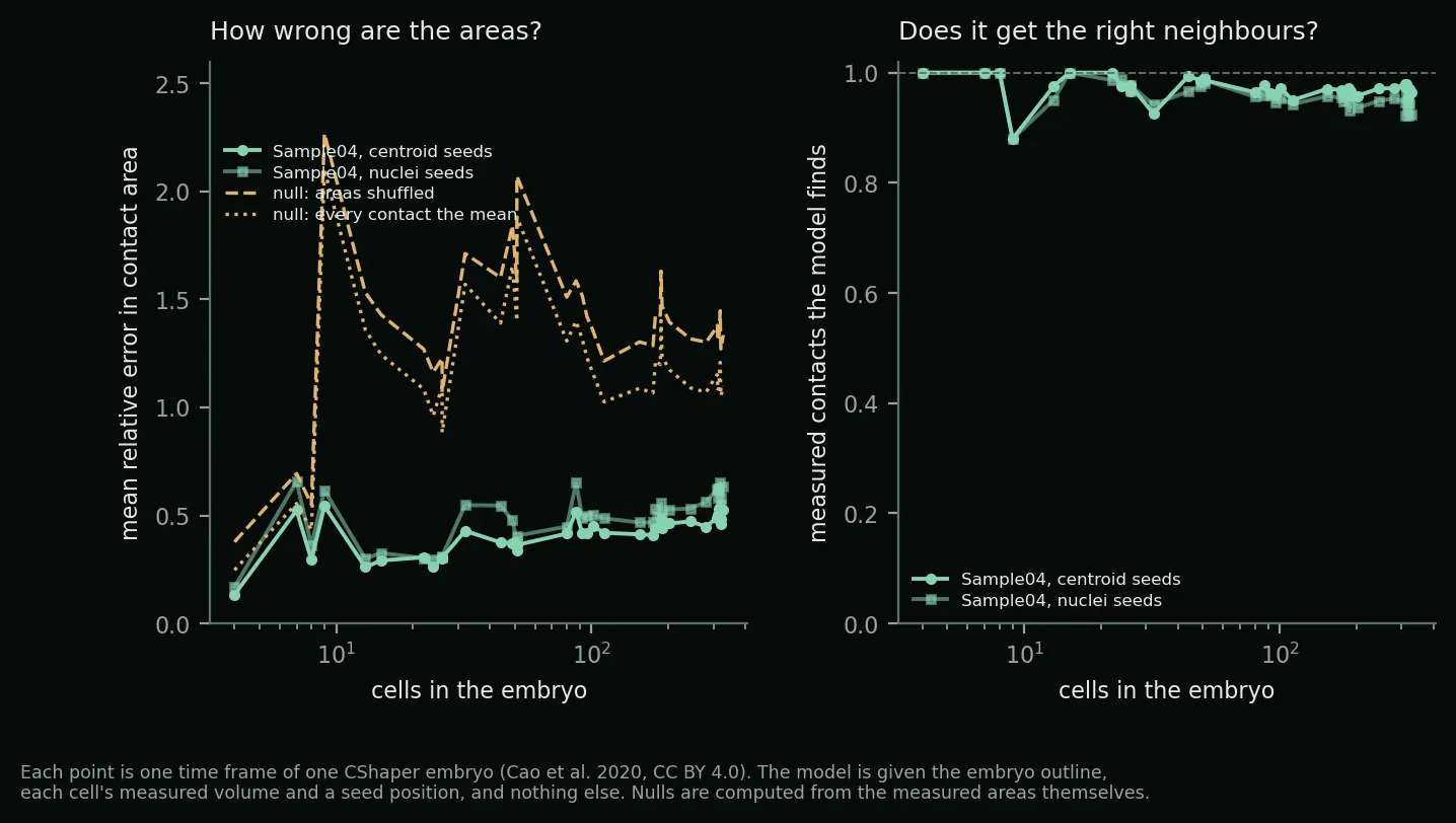
It finds 92–100% of the measured contacts at every stage from 4 to 328 cells. The areas are harder: within about 12% at four cells, drifting to about 50% at three hundred. To know whether that is any good we built two hard nulls — shuffle the measured areas among the measured pairs, and predict every contact at the frame's mean — and gave each of them the answer's own statistics. The model beats both in 38 frames out of 38. The page, with video.
07 · where the centres goThe field puts them in the wrong place
This one is simple, and it is the most immediately useful thing we found all day. Run the model over those thirty-eight frames twice, changing nothing but where each cell's centre is placed. Put it at the cell's measured centre of volume and the predicted contact areas land one to eleven points closer to the truth than putting it at the cell's nucleus — at every stage — and it finds more of the real contacts as well: 97% against 92% at three hundred cells.
That matters because predicting which cells touch from nuclear positions is the standard move in this field, and has been since the first whole-embryo reference model. Nuclei are what lineage tracing gives you. But a nucleus is not at the middle of its cell, and in a model where the centre is the cell's grip on space, that offset spreads into every wall it touches.
08 · borrowed from contract theoryIs a real embryo built the way we build ours?
Our embryo, and most others in the field, rests on one assumption: the wall between two cells is flat and square-on to the line joining their centres. It is made because it is convenient, and we could not find anyone who had put it to a measured embryo.
It can be checked without running anything. Give the arithmetic only the measured walls — no cell sizes, no nuclei, no lineage — and it works backwards to where the centres would have to be; rebuild the embryo from those and count how much returns. From four cells to three hundred and twenty, in two embryos, 92–100% of the real contacts return. Shuffle the walls between cells and almost none do.
Then the literature took most of the novelty away, and we think that is worth showing rather than hiding. A theorem from 1987, extended in 2008, says that any tessellation whose cells are convex and which meets face to face already is one of these diagrams. So the question worth asking is whether an embryo is convex and tidy — and it is, more than we expected. Cells fill eighty-nine to ninety-six per cent of their own convex hull, and past about eighty cells they are as convex as a diagram we built that way deliberately, because a real cell's rounded corners survive a voxel grid better than a mathematical one's sharp ones. Junctions where five cells meet are all but absent in both.
And the arithmetic we derived turns out to have been published in 2019 — in a paper about designing contracts that truthfully elicit what somebody believes. The same equations, for an entirely unrelated purpose.
Which, on reflection, is the interesting part. That particular paper has never been cited, in any field. The result it rests on has been cited 842 times without once reaching developmental biology: not one citing title mentions an embryo, morphogenesis, development, an epithelium or an organoid. Of its 29 biology citations, 23 are about packing atoms inside proteins — the tool crossed into biology and stopped three orders of magnitude short of a cell.
A correction to this section, made after a wider search. We first wrote that the tool and the problem had never met. That was too strong: the geometry reached economics properly, and the scoring-rules literature appears to have known since 2009. What never happened is the crossing into tissue-scale biology. The wider survey turned up something better than a simple gap anyway — fields holding the same object turn out to sit in several distinct states, including two cases of a field having the idea and then losing it. That survey is here.
What is ours: making that arithmetic survive noisy measurements — a case that literature never posed; giving the answer a floor and a chance level so a residual means something; and three numbers nobody had for an embryo — how convex its cells are, how flat its walls are (three per cent of their own width, the same at fifteen cells as at three hundred), and how far the remainder departs.
Two corrections, both made today. We first reported that the match weakens badly as the egg fills, and explained why. The weakening was ours: one number in the method was set a hundred times too strong, and it dragged the answer harder the more cells there were. We had even tested that decline against the dataset and cleared it — the test was sound for the question we asked, but it could not see a fault in the method rather than in the data. Then the prior-art search took the novelty of the arithmetic. Both are written up with the wrong values kept.
09 · two datasets with faultsThings we found in other people's data
The worm atlas has two traps. The C. elegans dataset we benchmark against stores voxel counts, not micrometres, and a second paper quantifying the same embryos converts surfaces and contacts by two different factors, explaining neither — its published areas come out 0.81× and 0.856× the first paper's. The deposit also numbers one kind of file from zero and every other kind from one, so the obvious join shifts every contact 1.39 minutes early. We verified the correct join on all 2,795 frames, against 859 for the naive one. Both groups are contactable and we intend to write to them.
Four embryos in a public transcriptome dataset are misdated. Labelled 32–35 hours after laying, they carry the whole-transcriptome profile of 5–12 hour embryos: excluding each other, their nearest neighbours are all early embryos, and a germline marker sits at its pre-clearance level. They fall exactly in the window our own expression tests depend on, so we reran every test without them — the verdicts stand, and one got cleaner. Nothing suggests intent; a label mix-up is the likeliest explanation, and it is flagged for the depositing lab.
10 · also todayAnd the rest
- Version 4 of the condensin II preprint is out. An independent recomputation reproduced every structure number but found the contact residue numbers were off by the length of a linker in the construct, and two claims worded too broadly. Both corrected; the errata are public.
- Six brain genes published with protein identifiers but no gene locations had never been joined to the expression data. They are now, with candidate locations for about fifty more neural markers — and two arrangements fell out that nobody has reported in a tardigrade: the two cholinergic genes sit as a tandem pair 5 kb apart, and three achaete-scute genes cluster within 17 kb, all three annotated only as "hypothetical protein".
- Nineteen open supplementary files fetched, including the only open 3D data of a stained tardigrade brain, each recorded with its licence and checksum.
- Velvet-worm mitochondrial genomes assembled overnight from public reads, in the separate onychophoran graph — the outgroup the tardigrade nervous system is always compared against.
- A muscle-placement fault, visible on the model. The routine that nudges leg muscles out of the organs was moving the left and right copies independently, and could squash or stretch a strand rather than move it. Leg four was worst, its muscles differing 36% between sides. Muscles are now moved as rigid bodies when nudging would deform them, and the build refuses to ship a left–right mismatch without reporting it.