Muscles through a different animal, and an embryo to hatching
We gave the glass tardigrade its leg muscles today: named from a colour code and fitted into this animal around its organs, from tracings Gross & Mayer made in another. Along the way the site split into a page per topic, everything we've built became downloadable, and in the afternoon the embryo grew to hatching.
A 4.9 GB zip from Dryad
We fetched the raw material behind Gross & Mayer's 2019 study of tardigrade leg muscles from Dryad: confocal stacks of all four legs and hand-traced label fields for every muscle. The first download turned out to be just their supplementary PDF, byte for byte a copy we already had. The real archive arrived second: 188 MB zipped, 4.9 GB unpacked.
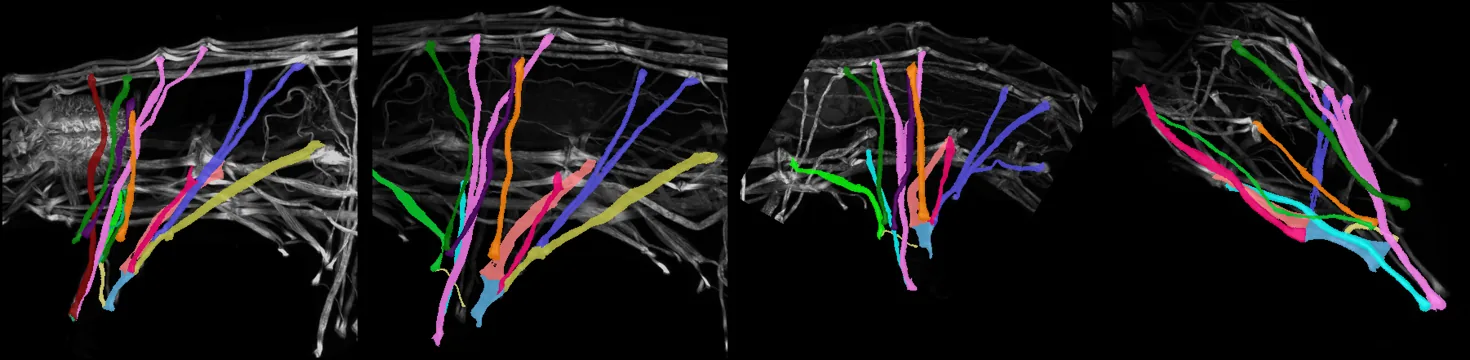
The colour code was the key
We named the muscles from their colours. The label files say "1, 2, 3", not "iM1, iM2", but the paper's Figure 2 colours each group of repeated muscles (M1 to M14) with its own colour, and the label files use the same palette. Counting colours per leg gave 14, 12, 11 and 10 muscles: exactly the paper's Table 1. Every label got its real name.
03 · a depth that didn't add up8 µm deep can't be right
The files claim 0.0425 µm per pixel in all three directions, which makes each leg only 8 µm thick. We asked what slice step makes the traced muscle strands round in cross-section. The answer was about 0.125 µm, the same in every leg. It's recorded as an estimate.
04 · one animal into anotherFitting the muscles
We pinned each leg's muscles at their claw end to that leg's claw gland in the X-ray animal, and at their top to its back wall, at one shared scale. The first try looked right from the side and wrong from below: the confocal stacks are squashed in depth under the coverslip, so the muscles sat in a thin sheet. Laying each muscle's depth inward from the X-ray animal's own body wall fixed that.
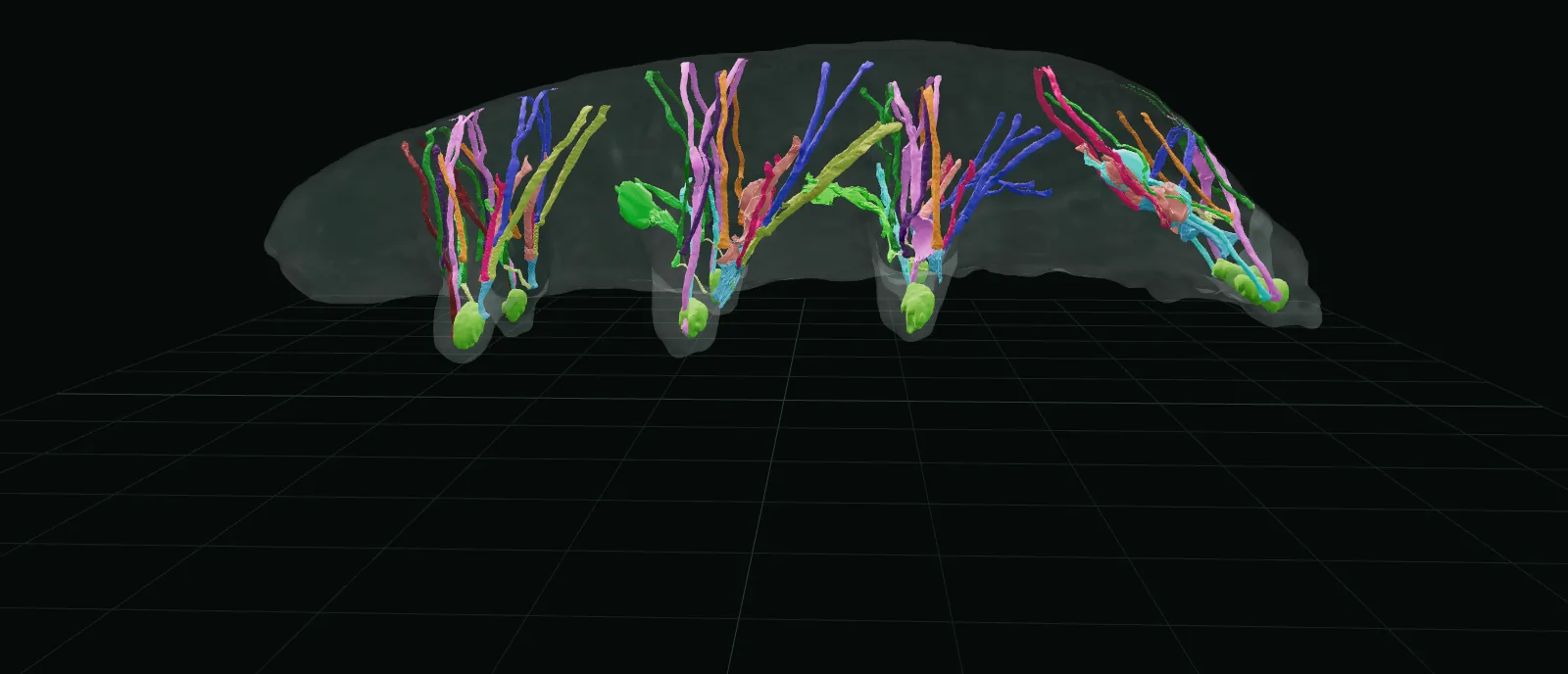
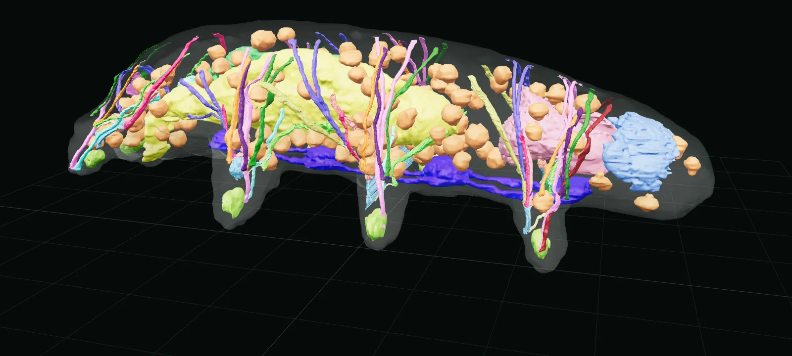
Muscles through storage cells
Meow-Ludo spotted muscles running straight through storage cells. They come from a different animal, whose cells were elsewhere. We marked this animal's storage cells, gut and ovary with a thin clearance, and pushed each piece of strand out along the shortest way, moving about a micrometre of strand at a time as a unit, so strands bend around a cell instead of splitting. 0.03% of muscle surface is left inside an organ: the slight clipping he said was fine.
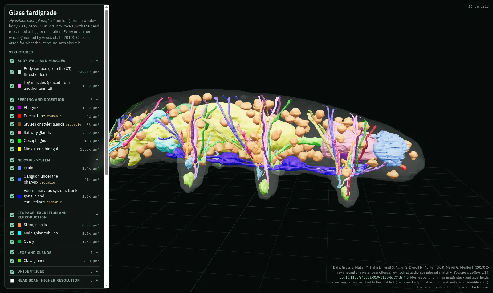
A grouped list
With twenty-one structures the list had grown long. It's now in groups (body wall and muscles, feeding and digestion, nervous system, storage, excretion and reproduction, legs and glands, unidentified, and the head scan), each collapsible and with a show-all checkbox.
A genome AI's tardigrade diet
A long download finished: the gene-focused windows of Evo 2's training data. Of 22.8 million windows, 2,481 are tardigrade and all but 37 come from one species, because only one tardigrade genome has the annotation format the pipeline used. Details on genomics.
08 · sharingPages per topic, and downloads
The site now has a page for each piece of work: the X-ray tomograph, muscles, walking, blocks and genomics. There's also a resources page with the organ and muscle meshes (GLB, STL, OBJ), a merged label volume with the head scan registered in, the body-masked CT, schematics and gait data, all CC BY 4.0, with a citation to copy.
09 · afternoon: the embryoFrom one cell to hatching
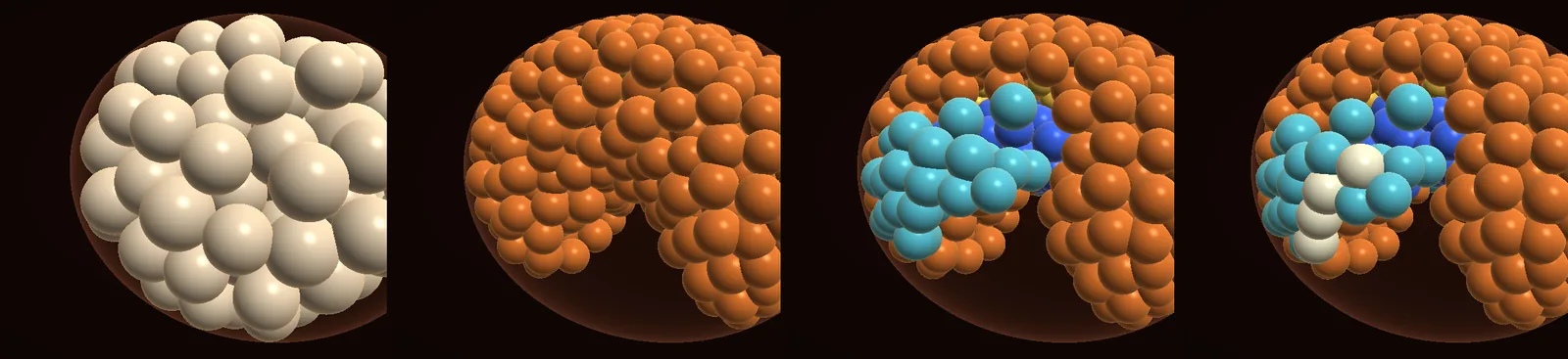
The afternoon went to the embryo. Two readers took in fifteen papers: gene expression in legs and segments, and the physics of cells packing in worm and sea urchin embryos. That added 144 claims and 26 open questions to the graph.
- Cell cycles: a new test compared the simulation cell by cell with Heikes' nine-embryo lineage. The first cycles were 15 minutes too slow; the measured ones now drive the embryo.
- Which way cells divide: cells now split along their longest free axis, after Pierre et al. 2016. Real divisions are recorded in how Heikes names cells, and this matches them more often than chance (64% against 59%), but not yet the real front-back bias (58% of divisions run front-back here against 71% in real embryos).
- Timing: seven sources, reconciled. The 2007 limb-bud time was about 10 hours early, and nothing else was.
- Stages 15–19: pharynx, ganglia, claws, eyespots, a roll in the shell and hatching are added. The ganglia came out largest at the front, as in adults. Cell counts at hatching are within about 5% of the 1,140 measured.
- A dataset's clock: Levin 2016's embryos match Yoshida's daily egg samples only on a ~98-hour axis. The paper's supplement has its minutes halved.
- Two bugs and a limit: cells moving inside had booked a 70-hour cycle, and gut cells stopped dividing after 21 hours. Both are fixed. The limb buds still won't separate, because spherical cells are too wide for four lobes.
The missing condensin piece was there after all
This morning we wrote that tardigrades have every condensin subunit except CAP-H2. BLAST couldn't find it, and that is the obvious gap a reviewer would press on. So we searched with a profile built from CAP-H2 in 33 other animals. Each of three tardigrades turns out to have two divergent copies. Each copy carries CAP-H2's signature domains and rises and falls through development with the rest of condensin II. The morning's wording is corrected on genomics. It also links up with an embryo imaging paper from 2026.