It hatched, and then the site grew to every tardigrade
The evening started with an embryo that gyrated in its shell and ended with a site about all 1,550 tardigrade species. In between, the embryo learned to hatch, we started giving every cell a cytoskeleton, and the site gained a tree of every species, a gallery of 415 figures from the literature, and pages on the brain, drying out and surviving the extremes.
From a bunch of spheres to an animal
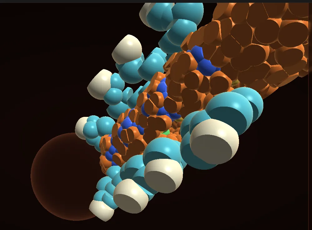
Meow-Ludo's screenshots showed the late embryo gyrating and looking like a pile of balls. Gabriel's time-lapse movies showed what was wrong: a real embryo fills its shell from the first cell to hatching, its skin is smooth, and it only turns now and then. The stylets in one movie hold still for about 13 hours at a stretch. So the embryo now turns occasionally instead of spinning, fills the shell, closes its skin over the growing ganglia, and has rounded ends and legs that point down.
It hatched
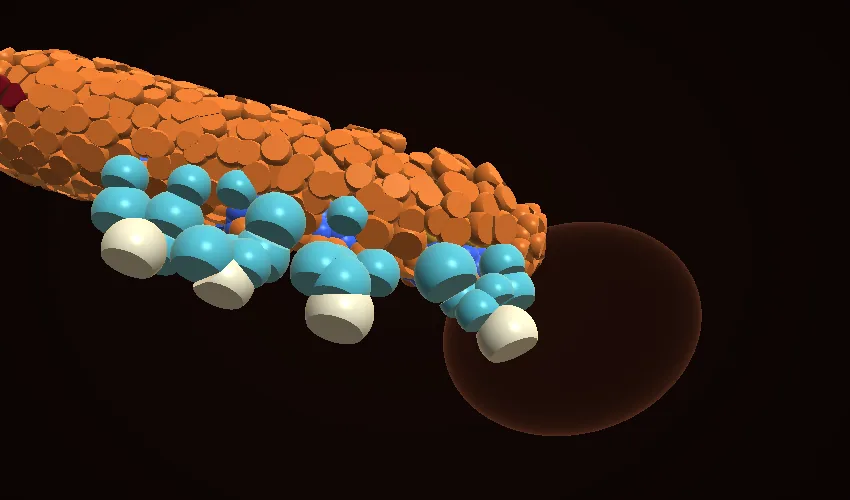
The embryo had been running on past hatching, and its cells kept dividing: 1,700 and counting. Now, at 102 hours, the stylets open a hole at the head end, and the animal slides out along its own curl, straightens and lengthens beside the empty shell, the way the 2007 description and movie show it. The clock stops once it's out, at about 1,090 cells against 1,140 counted in real hatchlings. A top bar now shows the cell count, the stage and a progress bar to hatching.
Every cell a small graph
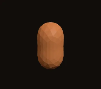
Painting shapes onto points was never going to make columnar skin or separate limb buds. So each cell is becoming a small graph: a membrane of a few hundred points around a centre node, joined by spokes that stand for its microtubules, with a tensor that sets its main axes. Volume stays fixed, the cortex pulls the surface tight, and neighbours stick where they touch. One strong axis turns a cell into a column.
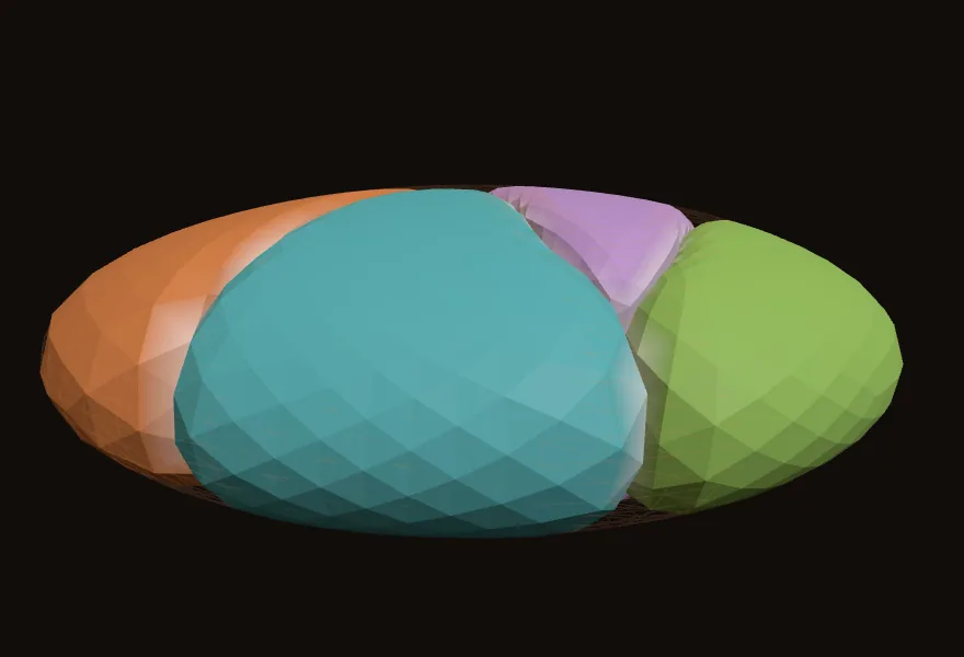
We tested it on the best-measured embryo there is. With the worm's measured cell volumes and division axes in its flattened eggshell, four cells settle into the real rhomboid, with all five contacts and none between ABa and P2. The error on contact and surface areas is 0.18, against 0.06 for the best published model; our contacts are still about 30% small. Getting there took several fixes: cells that started inside each other, walls that crumpled into flaps, a shell squashed along the wrong axis, and a units mix-up in the measured areas that we traced to how voxel faces overestimate curved surfaces.
1,550 species, one tree
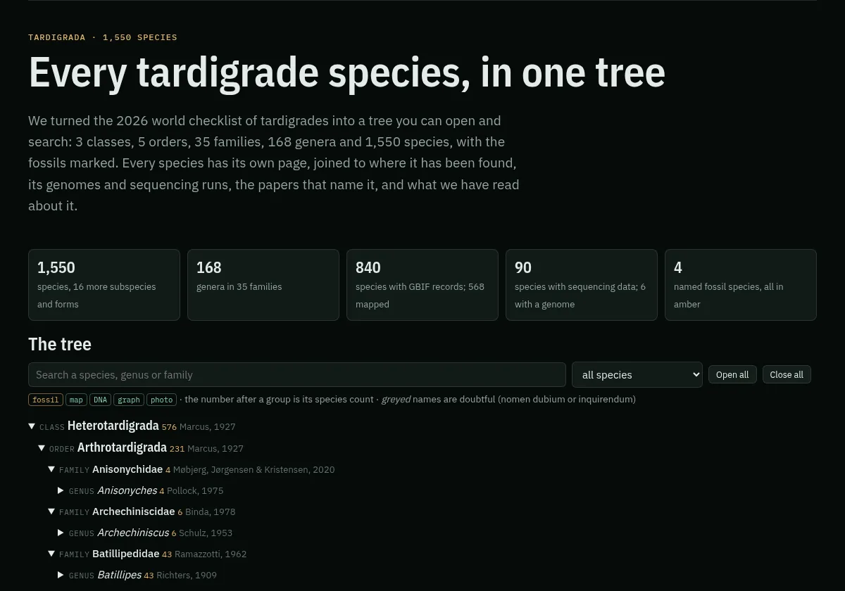
We turned the 2026 world checklist into a tree of 3 classes, 5 orders, 35 families, 168 genera and 1,550 species, with the four fossil species in amber marked. Every species has its own page: where it has been found, its sequencing data, how many papers name it, openly licensed photos, and anything our graph knows about it.
415 figures, credited
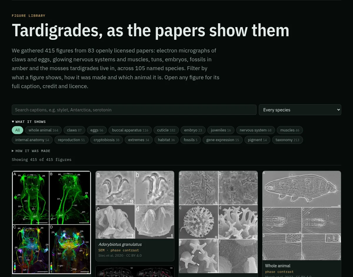
Tardigrade papers are full of beautiful images. We gathered 415 from 83 openly licensed papers: electron micrographs of claws and eggs, glowing nervous systems and muscles, tuns, embryos and the mosses they live in. Each keeps its full caption, credit and licence, and the gallery filters by what a figure shows, how it was made and which species. Topic pages now pull in matching figures.
The brain, drying out, and the real extremes
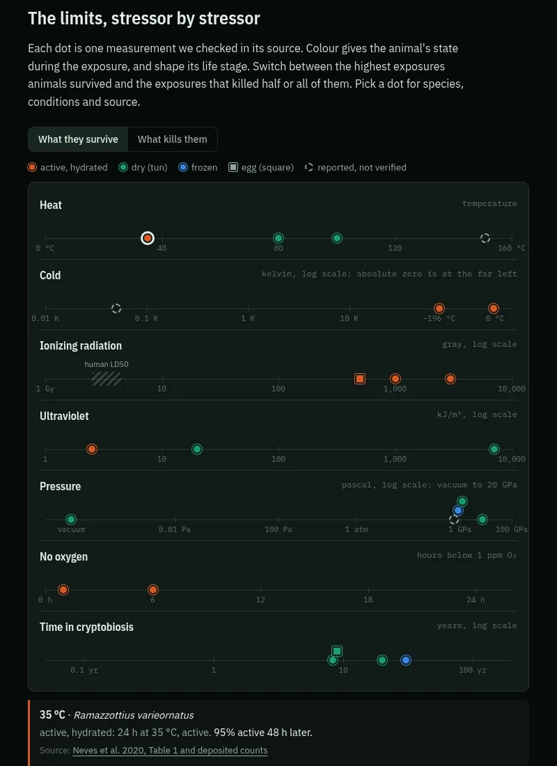
The brain page has a clickable map of the nervous system, shown two ways: by structure, and by the open questions. It also has new measurements of ours from the scan: the trunk ganglia sit 4.6–12.2 µm in front of their legs, with volumes of about 160–410 µm³. Cryptobiosis covers drying, freezing and the protective proteins. Extremes traces every famous survival figure back to its source: the species, whether the animal was active or dried, and the exact conditions. The heat curves are redrawn from the raw counts one lab deposited, and eight popular claims are checked, from "near absolute zero" to the Moon crash.
And the rest
- Find and keep tardigrades: collecting from moss, the story of the lab strain (one female from a Bolton pond in 1987), culture recipes and diets.
- Open questions: 228 questions, 145 quoted from the papers that asked them, with the answers later work has found.
- Findings, every public dataset, a history from 1773 to today, and an animated life cycle.
- Condensin II turned out to be complete in tardigrades: two divergent copies of the missing subunit, found by profile search (genomics).
- The menu is now grouped, and several pages offer another view of their data: charts beside tables, a board beside the list of findings, and questions by theme or by paper.