From label files to a walking glass animal
In two days we found published scans, tracings, films and counts of one species, worked out what they meant, and joined them in one frame: a glass tardigrade you can cut open, a walking one, and a Minecraft build. The measurements are the published work of the scientists credited at each step.
A whole water bear, scanned by X-ray
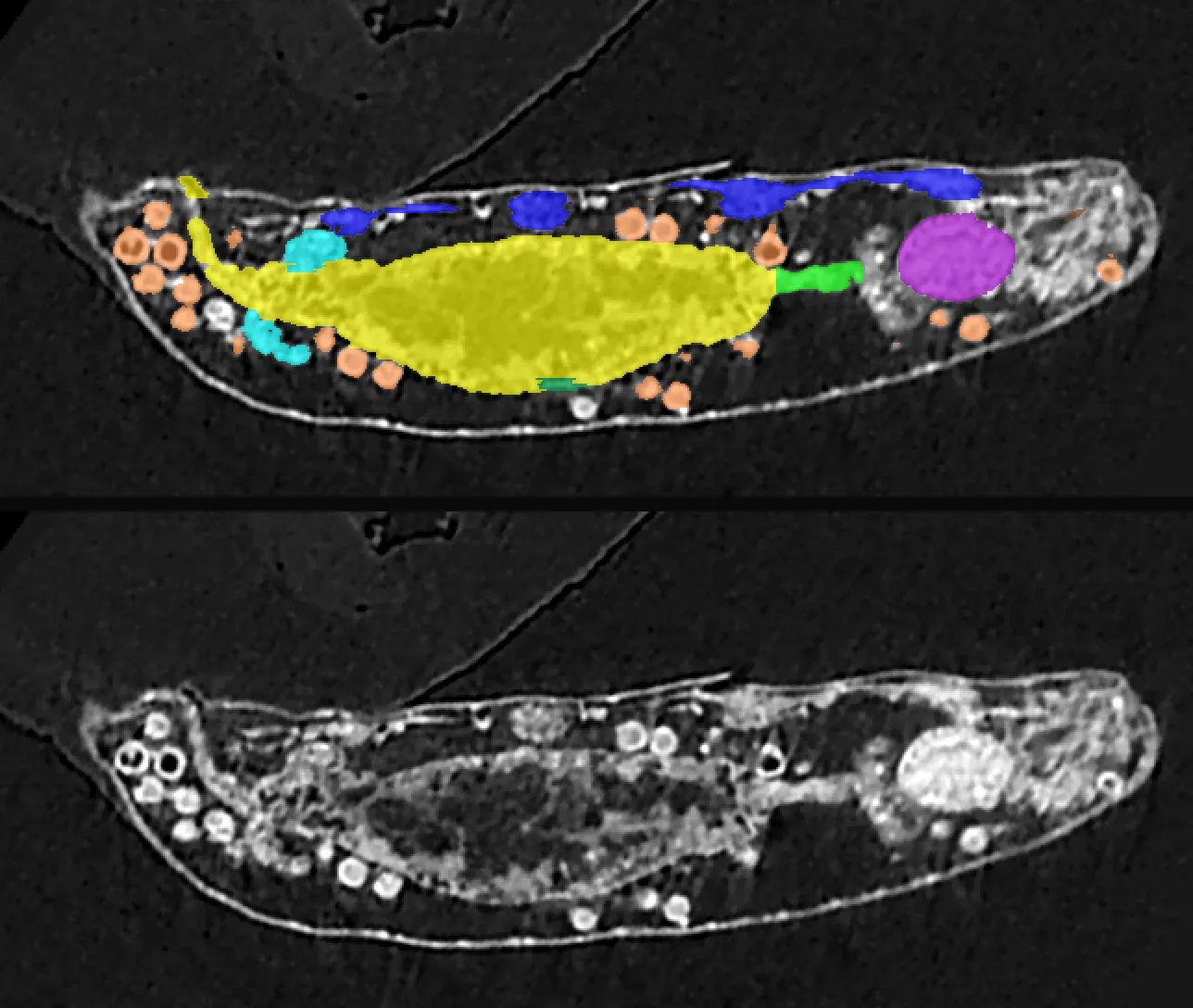
We started from an adult Hypsibius exemplaris scanned whole in a lab nano-CT at 270 nm per voxel, its head rescanned at higher resolution and every organ traced by hand, slice by slice. Vladimir Gross, Georg Mayer, Franz Pfeiffer and colleagues published the image stacks and the tracing ("label fields") openly in 2019, under CC BY.
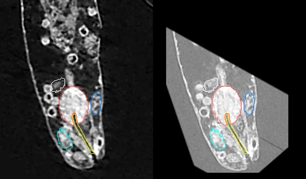
The label files hold numbers, not names: 1, 2, 3 up to 11. We measured every label and matched it to the organ volumes in the paper's Table 1: label 1 is 1,763 µm³ against the pharynx's published 1,771; label 3 is 13,812 against the gut's 13,706. Two labels sharing one colour in the file's palette turned out to be the Malpighian tubules, together within 1% of the published volume.
Meshes, and a body that wasn't labelled
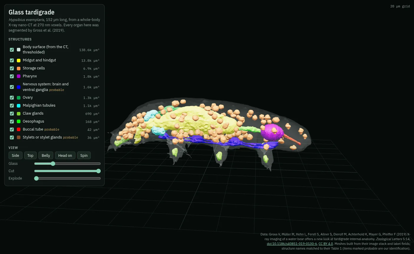
Each label became a surface with marching cubes (the standard way to wrap a skin around voxels), then simplified and packed into one 3D file for the browser. The body itself had no label, so we traced it from the bright rim of the cuticle in the CT and filled it in, landing within 1% of the published body volume.
The webbing between the legs
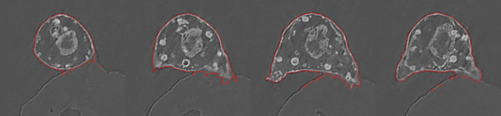
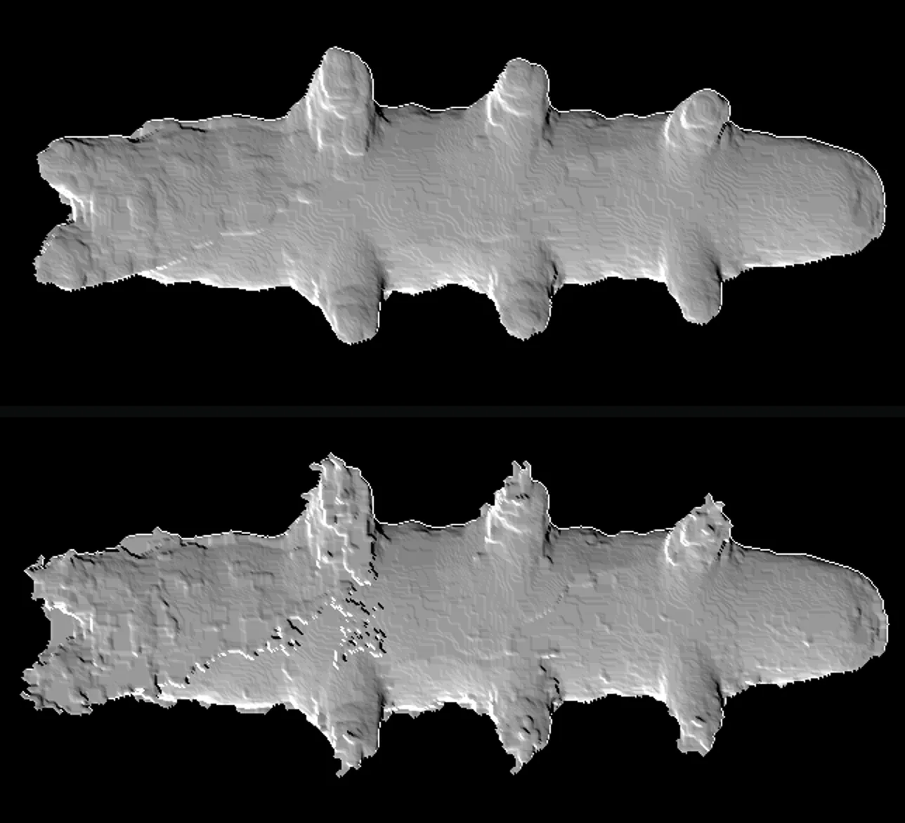
Meow-Ludo spotted extra skin between the hind legs. The CT showed why: the animal was lying against a thin film and a curved holder, and those closed off pockets beside its legs, which the fill step dutifully filled. The fix was to keep a voxel only when two of three slicing directions agree it's inside, then erase sheets thinner than 1.6 µm. The body volume dropped to 137,068 µm³, within 0.3% of the paper's 137,415.
The head scan, and a size that didn't add up
We brought in the separate head scan, the only place the brain and salivary glands were traced. The paper says that scan used 200 nm voxels, but at that size the organs came out far too big. At 0.1065 µm per voxel, two independent organs matched the paper exactly: brain 1,389 µm³ against 1,393, salivary glands 2,274 against 2,274.
To place the head inside the body, we matched the two X-ray volumes directly, trying all 24 ways a cube can be turned and sliding each over the other with a fast Fourier correlation. One orientation won clearly. Afterwards 87% of the head scan's pharynx sat inside the whole-body pharynx, and a ganglion under the pharynx lined up with the nerve cord.
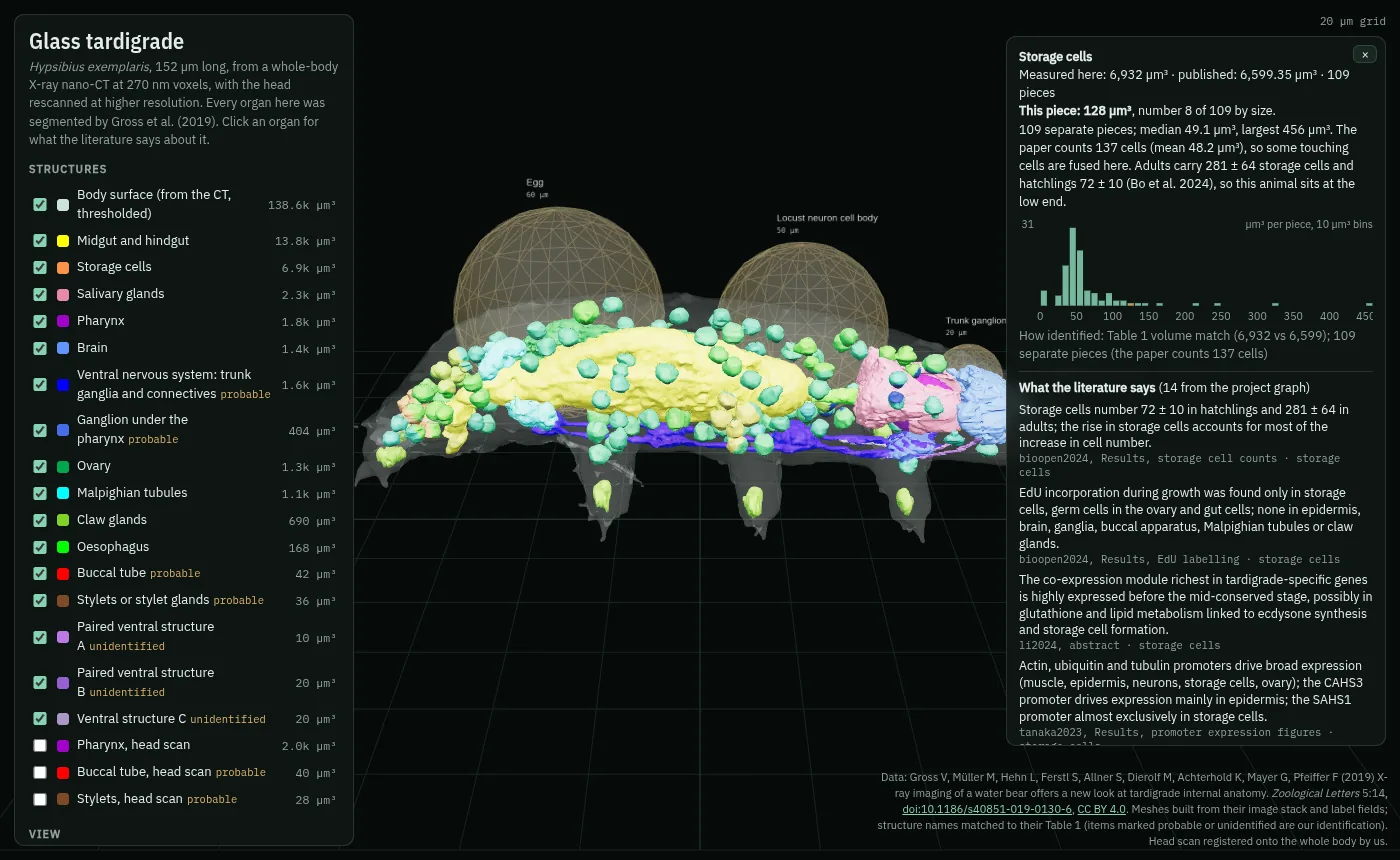
Cut it open, count its cells
The cut plane samples the real CT, so slicing the glass animal shows the X-ray at that position. The 109 storage cells are individually clickable, with a histogram of their volumes. The scan's 137 storage cells sit between a hatchling's 72 and an adult's 281, fitting a 152 µm animal that was still growing.
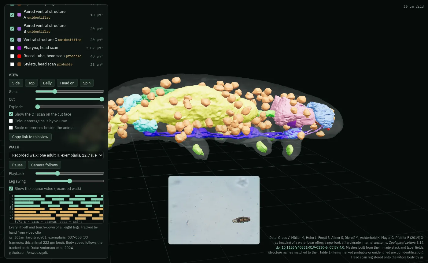
Making it walk
We made it walk to footsteps that Ian Woods' lab marked by hand in video, every touch-down and lift-off, shared with their code and spreadsheets on GitHub. We found each foot on the CT animal from its claw gland, bent the skin around each leg, and played back their recording step for step, next to the original video.
Left and right mattered. The scan has no labels for sides, so we checked the published anatomy: the ovary lies on the animal's left, which fixed which legs were which.
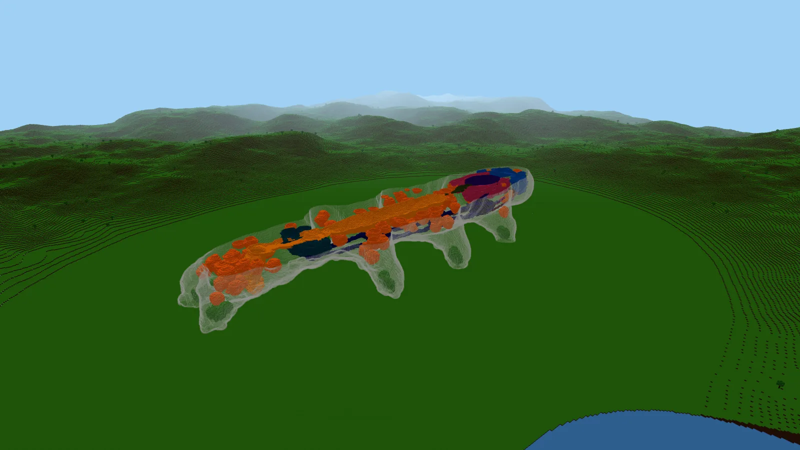
A tardigrade in Minecraft
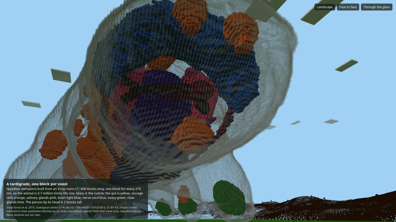
A voxel is already a block. One block per 270 nm voxel makes the animal 608 blocks long and 3.7 million times life size. The skin is one layer of glass; organs are coloured concrete; the head scan's brain and salivary glands are placed the same way as in the glass viewer. The build exports as a schematic you can paste into a Minecraft world.
The X-ray, in stone
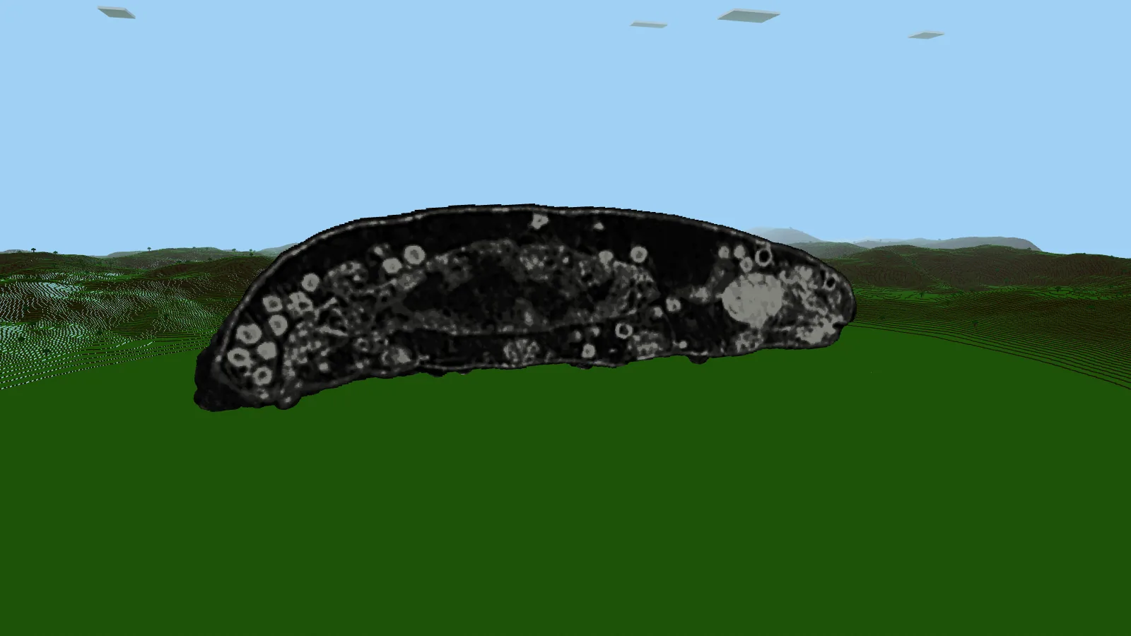
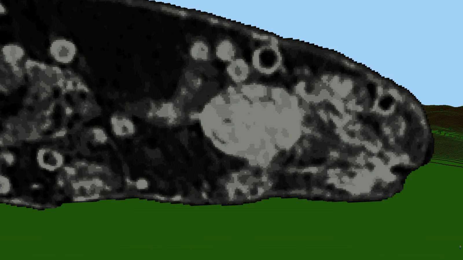
The same trick works on the raw scan. Every voxel inside the body becomes one of eleven grey blocks: black concrete, polished blackstone, deepslate, tuff, stone, light grey concrete, andesite, smooth stone, diorite, calcite and quartz. A straight brightness scale turned nearly everything black, because the body cavity is almost the grey of the liquid around it. So the stones are spread halfway between equal counts and a straight scale. Cut down the middle, the storage cells show as pale rings and the pharynx as a bright ball.
The embryo
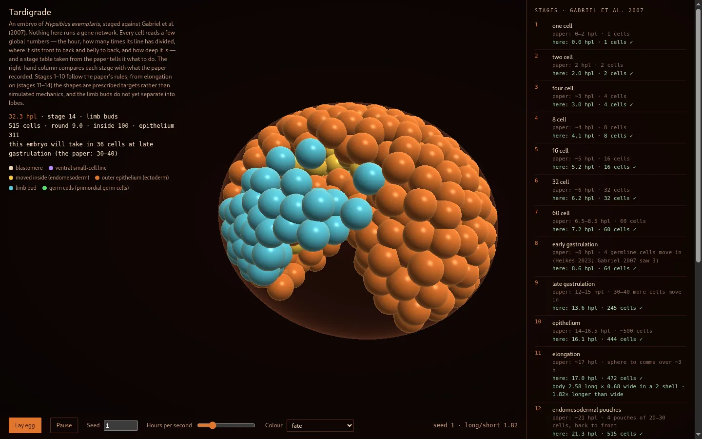
Separately, we grew an embryo cell by cell against the 2007 staging series of Willow Gabriel, Bob Goldstein and colleagues: the hours and cell counts for each stage sit beside the simulation as it runs. It now follows the four cells that become the germline inside, as Kira Heikes and colleagues traced them in 2023.
What's next
Muscles from confocal scans of the legs, a named nervous system, the animal moulting, and the same method on other tardigrade species. Every step will stay traceable to the people who measured it.
Full list of data and licences: credits.